2J3N

X-ray structure of human thioredoxin reductase 1


Experimental Data Snapshot

  • Method: X-RAY DIFFRACTION
  • Resolution: 2.80 Å
  • R-Value Free: 0.287 
  • R-Value Work: 0.249 
  • R-Value Observed: 0.249 

wwPDB Validation   3D Report Full Report


Ligand Structure Quality Assessment 


This is version 1.4 of the entry. See complete history


Literature

The Structure of Human Thioredoxin Reductase 1 Provides Insights Into C-Terminal Rearrangements During Catalysis.

Fritz-Wolf, K.Urig, S.Becker, K.

(2007) J Mol Biol 370: 116

  • DOI: https://doi.org/10.1016/j.jmb.2007.04.044
  • Primary Citation of Related Structures:  
    2J3N

  • PubMed Abstract: 

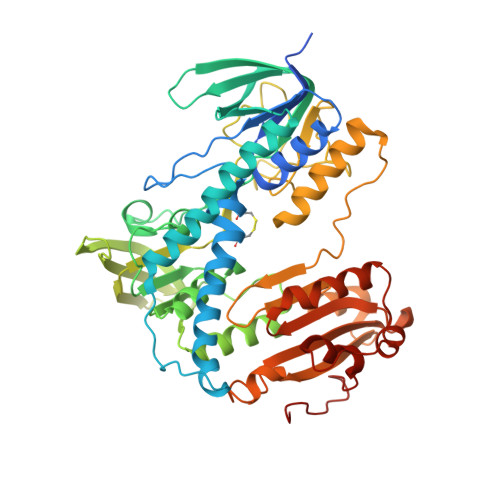
    Human thioredoxin reductase (hTrxR) is a homodimeric flavoprotein crucially involved in the regulation of cellular redox reactions, growth and differentiation. The enzyme contains a selenocysteine residue at its C-terminal active site that is essential for catalysis. This redox center is located on a flexible arm, solvent-exposed and reactive towards electrophilic inhibitors, thus representing a target for antitumor drug development. During catalysis reducing equivalents are transferred from the cofactor NADPH to FAD, then to the N-terminal active site cysteine residues and from there to the flexible C-terminal part of the other subunit to be finally delivered to a variety of second substrates at the molecule's surface. Here we report the first crystal structure of hTrxR1 (Sec-->Cys) in complex with FAD and NADP(+) at a resolution of 2.8 A. From the crystals three different conformations of the carboxy-terminal arm could be deduced. The predicted movement of the arm is facilitated by the concerted action of the three side-chain residues of N418, N419 and W407, which act as a guiding bar for the C-terminal sliding process. As supported by previous kinetic data, the three visualized conformations might reflect different stages in enzymatic catalysis. Comparison with other disulfide reductases including human glutathione reductase revealed specific inhibitor binding sites in the intersubunit cavity of hTrxR that can be exploited for structure-based inhibitor development.


  • Organizational Affiliation

    Interdisciplinary Research Center, Justus-Liebig-University, 35392 Giessen, Germany.


Macromolecules
Find similar proteins by:  (by identity cutoff)  |  3D Structure
Entity ID: 1
MoleculeChains Sequence LengthOrganismDetailsImage
THIOREDOXIN REDUCTASE 1
A, B, C, D, E
A, B, C, D, E, F
519Homo sapiensMutation(s): 0 
EC: 1.8.1.9
UniProt & NIH Common Fund Data Resources
Find proteins for Q16881 (Homo sapiens)
Explore Q16881 
Go to UniProtKB:  Q16881
PHAROS:  Q16881
GTEx:  ENSG00000198431 
Entity Groups  
Sequence Clusters30% Identity50% Identity70% Identity90% Identity95% Identity100% Identity
UniProt GroupQ16881
Sequence Annotations
Expand
  • Reference Sequence
Small Molecules
Ligands 3 Unique
IDChains Name / Formula / InChI Key2D Diagram3D Interactions
FAD
Query on FAD

Download Ideal Coordinates CCD File 
G [auth A]
J [auth B]
N [auth C]
P [auth D]
S [auth E]
G [auth A],
J [auth B],
N [auth C],
P [auth D],
S [auth E],
U [auth F]
FLAVIN-ADENINE DINUCLEOTIDE
C27 H33 N9 O15 P2
VWWQXMAJTJZDQX-UYBVJOGSSA-N
NAP
Query on NAP

Download Ideal Coordinates CCD File 
H [auth A]
K [auth B]
O [auth C]
Q [auth D]
T [auth E]
H [auth A],
K [auth B],
O [auth C],
Q [auth D],
T [auth E],
V [auth F]
NADP NICOTINAMIDE-ADENINE-DINUCLEOTIDE PHOSPHATE
C21 H28 N7 O17 P3
XJLXINKUBYWONI-NNYOXOHSSA-N
MPD
Query on MPD

Download Ideal Coordinates CCD File 
I [auth A],
L [auth B],
M [auth B],
R [auth D],
W [auth F]
(4S)-2-METHYL-2,4-PENTANEDIOL
C6 H14 O2
SVTBMSDMJJWYQN-YFKPBYRVSA-N
Experimental Data & Validation

Experimental Data

  • Method: X-RAY DIFFRACTION
  • Resolution: 2.80 Å
  • R-Value Free: 0.287 
  • R-Value Work: 0.249 
  • R-Value Observed: 0.249 
  • Space Group: P 1 21 1
Unit Cell:
Length ( Å )Angle ( ˚ )
a = 144.63α = 90
b = 90.64β = 112.46
c = 166.6γ = 90
Software Package:
Software NamePurpose
CNSrefinement
XDSdata reduction
XDSdata scaling
CNSphasing

Structure Validation

View Full Validation Report


Ligand Structure Quality Assessment 


Entry History 

Deposition Data

Revision History  (Full details and data files)

  • Version 1.0: 2007-07-03
    Type: Initial release
  • Version 1.1: 2011-05-08
    Changes: Version format compliance
  • Version 1.2: 2011-07-13
    Changes: Version format compliance
  • Version 1.3: 2018-01-24
    Changes: Source and taxonomy
  • Version 1.4: 2023-12-13
    Changes: Data collection, Database references, Derived calculations, Other, Refinement description