2IJ7

Structure of Mycobacterium tuberculosis CYP121 in complex with the antifungal drug fluconazole


Experimental Data Snapshot

  • Method: X-RAY DIFFRACTION
  • Resolution: 1.90 Å
  • R-Value Free: 0.226 
  • R-Value Work: 0.167 
  • R-Value Observed: 0.170 

wwPDB Validation   3D Report Full Report


Ligand Structure Quality Assessment 


This is version 1.3 of the entry. See complete history


Literature

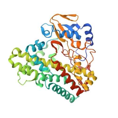
Crystal structure of the Mycobacterium tuberculosis P450 CYP121-fluconazole complex reveals new azole drug-P450 binding mode.

Seward, H.E.Roujeinikova, A.McLean, K.J.Munro, A.W.Leys, D.

(2006) J Biol Chem 281: 39437-39443

  • DOI: https://doi.org/10.1074/jbc.M607665200
  • Primary Citation of Related Structures:  
    2IJ5, 2IJ7

  • PubMed Abstract: 

    Azole and triazole drugs are cytochrome P450 inhibitors widely used as fungal antibiotics and possessing potent antimycobacterial activity. We present here the crystal structure of Mycobacterium tuberculosis cytochrome P450 CYP121 in complex with the triazole drug fluconazole, revealing a new azole heme ligation mode. In contrast to other structurally characterized cytochrome P450 azole complexes, where the azole nitrogen directly coordinates the heme iron, in CYP121 fluconazole does not displace the aqua sixth heme ligand but occupies a position that allows formation of a direct hydrogen bond to the aqua sixth heme ligand. Direct ligation of fluconazole to the heme iron is observed in a minority of CYP121 molecules, albeit with severe deviations from ideal geometry due to close contacts with active site residues. Analysis of both ligand-on and -off structures reveals the relative position of active site residues derived from the I-helix is a key determinant in the relative ratio of on and off states. Regardless, both ligand-bound states lead to P450 inactivation by active site occlusion. This previously unrecognized means of P450 inactivation is consistent with spectroscopic analyses in both solution and in the crystalline form and raises important questions relating to interaction of azoles with both pathogen and human P450s.


  • Organizational Affiliation

    Department of Biochemistry, University of Leicester, Henry Wellcome Building, Lancaster Road, Leicester LE6 0HQ, United Kingdom.


Macromolecules
Find similar proteins by:  (by identity cutoff)  |  3D Structure
Entity ID: 1
MoleculeChains Sequence LengthOrganismDetailsImage
Cytochrome P450 121
A, B, C, D, E
A, B, C, D, E, F
396Mycobacterium tuberculosisMutation(s): 0 
Gene Names: cyp121
EC: 1.14
UniProt
Find proteins for P9WPP7 (Mycobacterium tuberculosis (strain ATCC 25618 / H37Rv))
Explore P9WPP7 
Go to UniProtKB:  P9WPP7
Entity Groups  
Sequence Clusters30% Identity50% Identity70% Identity90% Identity95% Identity100% Identity
UniProt GroupP9WPP7
Sequence Annotations
Expand
  • Reference Sequence
Small Molecules
Ligands 2 Unique
IDChains Name / Formula / InChI Key2D Diagram3D Interactions
HEM
Query on HEM

Download Ideal Coordinates CCD File 
G [auth A]
I [auth B]
K [auth C]
M [auth D]
O [auth E]
G [auth A],
I [auth B],
K [auth C],
M [auth D],
O [auth E],
P [auth F]
PROTOPORPHYRIN IX CONTAINING FE
C34 H32 Fe N4 O4
KABFMIBPWCXCRK-RGGAHWMASA-L
TPF
Query on TPF

Download Ideal Coordinates CCD File 
H [auth A],
J [auth B],
L [auth C],
N [auth D],
Q [auth F]
2-(2,4-DIFLUOROPHENYL)-1,3-DI(1H-1,2,4-TRIAZOL-1-YL)PROPAN-2-OL
C13 H12 F2 N6 O
RFHAOTPXVQNOHP-UHFFFAOYSA-N
Binding Affinity Annotations 
IDSourceBinding Affinity
TPF BindingDB:  2IJ7 Kd: 6000 (nM) from 1 assay(s)
Binding MOAD:  2IJ7 Kd: 5000 (nM) from 1 assay(s)
Experimental Data & Validation

Experimental Data

  • Method: X-RAY DIFFRACTION
  • Resolution: 1.90 Å
  • R-Value Free: 0.226 
  • R-Value Work: 0.167 
  • R-Value Observed: 0.170 
  • Space Group: P 21 21 21
Unit Cell:
Length ( Å )Angle ( ˚ )
a = 60.805α = 90
b = 129.898β = 90
c = 323.393γ = 90
Software Package:
Software NamePurpose
REFMACrefinement
ADSCdata collection
DENZOdata reduction
SCALEPACKdata scaling
PHASERphasing

Structure Validation

View Full Validation Report


Ligand Structure Quality Assessment 


Entry History 

Deposition Data

Revision History  (Full details and data files)

  • Version 1.0: 2006-11-07
    Type: Initial release
  • Version 1.1: 2008-05-01
    Changes: Version format compliance
  • Version 1.2: 2011-07-13
    Changes: Version format compliance
  • Version 1.3: 2023-08-30
    Changes: Data collection, Database references, Derived calculations, Refinement description