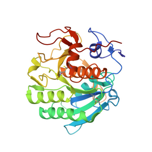
2ID8

Crystal structure of Proteinase K


Experimental Data Snapshot

  • Method: X-RAY DIFFRACTION
  • Resolution: 1.27 Å
  • R-Value Free: 0.159 
  • R-Value Observed: 0.124 

wwPDB Validation   3D Report Full Report


Ligand Structure Quality Assessment 


This is version 1.2 of the entry. See complete history


Literature

What can be done with a good crystal and an accurate beamline?

Wang, J.Dauter, M.Dauter, Z.

(2006) Acta Crystallogr D Biol Crystallogr 62: 1475-1483

  • DOI: https://doi.org/10.1107/S0907444906038534
  • Primary Citation of Related Structures:  
    2ID8

  • PubMed Abstract: 

    X-ray single-wavelength anomalous diffraction (SAD) data from a crystal of proteinase K were collected using synchrotron radiation of 0.98 A wavelength at SER-CAT 22-ID beamline, Advanced Photon Source, Argonne National Laboratory. At this wavelength, the expected Bijvoet ratio resulting from the presence of one calcium, one chloride and ten S atoms in the 279-residue protein is extremely small at approximately 0.46%. The direct-methods program SHELXD located 11 anomalous sites using data truncated to 2 A resolution. SHELXE was used to produce an easily interpretable electron-density map. This study shows that an accurate beamline and a good-quality crystal provide the possibility of successfully using a very weak anomalous signal of sulfur measured at a short wavelength for phasing a protein structure, even if a small degree of radiation damage is present.


  • Organizational Affiliation

    SAIC-Frederick Inc., Basic Research Program, Argonne National Laboratory, Argonne, IL 60439, USA.


Macromolecules
Find similar proteins by:  (by identity cutoff)  |  3D Structure
Entity ID: 1
MoleculeChains Sequence LengthOrganismDetailsImage
Proteinase K279Parengyodontium albumMutation(s): 1 
EC: 3.4.21.64
UniProt
Find proteins for P06873 (Parengyodontium album)
Explore P06873 
Go to UniProtKB:  P06873
Entity Groups  
Sequence Clusters30% Identity50% Identity70% Identity90% Identity95% Identity100% Identity
UniProt GroupP06873
Sequence Annotations
Expand
  • Reference Sequence
Experimental Data & Validation

Experimental Data

  • Method: X-RAY DIFFRACTION
  • Resolution: 1.27 Å
  • R-Value Free: 0.159 
  • R-Value Observed: 0.124 
  • Space Group: P 43 21 2
Unit Cell:
Length ( Å )Angle ( ˚ )
a = 67.55α = 90
b = 67.55β = 90
c = 106.88γ = 90
Software Package:
Software NamePurpose
SHELXmodel building
SHELXL-97refinement
SERGUIdata collection
HKL-2000data reduction
HKL-2000data scaling
SHELXDphasing
SHELXEmodel building

Structure Validation

View Full Validation Report


Ligand Structure Quality Assessment 


Entry History 

Deposition Data

Revision History  (Full details and data files)

  • Version 1.0: 2006-10-10
    Type: Initial release
  • Version 1.1: 2008-05-01
    Changes: Version format compliance
  • Version 1.2: 2011-07-13
    Changes: Version format compliance