


Crystal structure of an adenine deaminase
Sugadev, R., Kumaran, D., Burley, S.K., Swaminathan, S.To be published.
Experimental Data Snapshot
wwPDB Validation 3D Report Full Report

Entity ID: 1 | |||||
|---|---|---|---|---|---|
| Molecule | Chains | Sequence Length | Organism | Details | Image |
| Adenine Deaminase | 379 | Enterococcus faecalis | Mutation(s): 7 EC: 3.5.4.2 | 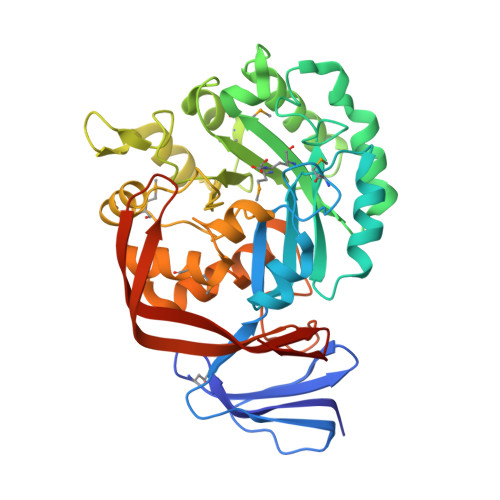 | |
UniProt | |||||
Find proteins for Q837K0 (Enterococcus faecalis (strain ATCC 700802 / V583)) Explore Q837K0 Go to UniProtKB: Q837K0 | |||||
Entity Groups | |||||
| Sequence Clusters | 30% Identity50% Identity70% Identity90% Identity95% Identity100% Identity | ||||
| UniProt Group | Q837K0 | ||||
Sequence AnnotationsExpand | |||||
| |||||
| Ligands 2 Unique | |||||
|---|---|---|---|---|---|
| ID | Chains | Name / Formula / InChI Key | 2D Diagram | 3D Interactions | |
| ADE Query on ADE | D [auth A] | ADENINE C5 H5 N5 GFFGJBXGBJISGV-UHFFFAOYSA-N |  | ||
| ZN Query on ZN | B [auth A], C [auth A] | ZINC ION Zn PTFCDOFLOPIGGS-UHFFFAOYSA-N |  | ||
| Modified Residues 2 Unique | |||||
|---|---|---|---|---|---|
| ID | Chains | Type | Formula | 2D Diagram | Parent |
| KCX Query on KCX | A | L-PEPTIDE LINKING | C7 H14 N2 O4 |  | LYS |
| MSE Query on MSE | A | L-PEPTIDE LINKING | C5 H11 N O2 Se |  | MET |
| Length ( Å ) | Angle ( ˚ ) |
|---|---|
| a = 62.373 | α = 90 |
| b = 141.686 | β = 90 |
| c = 47.396 | γ = 90 |
| Software Name | Purpose |
|---|---|
| CNS | refinement |
| CBASS | data collection |
| HKL-2000 | data reduction |
| HKL-2000 | data scaling |
| SHELXD | phasing |
| SHARP | phasing |
| ARP/wARP | model building |
RCSB PDB (citation) is hosted by
RCSB PDB is a member of the

