2IBY

Crystallographic and kinetic studies of human mitochondrial acetoacetyl-CoA thiolase (T2): the importance of potassium and chloride for its structure and function


Experimental Data Snapshot

  • Method: X-RAY DIFFRACTION
  • Resolution: 1.85 Å
  • R-Value Free: 0.203 
  • R-Value Work: 0.164 
  • R-Value Observed: 0.166 

wwPDB Validation   3D Report Full Report


Ligand Structure Quality Assessment 


This is version 1.5 of the entry. See complete history


Literature

Crystallographic and Kinetic Studies of Human Mitochondrial Acetoacetyl-CoA Thiolase: The Importance of Potassium and Chloride Ions for Its Structure and Function

Haapalainen, A.M.Merilainen, G.Pirila, P.L.Kondo, N.Fukao, T.Wierenga, R.K.

(2007) Biochemistry 46: 4305-4321

  • DOI: https://doi.org/10.1021/bi6026192
  • Primary Citation of Related Structures:  
    2IB7, 2IB8, 2IB9, 2IBU, 2IBW, 2IBY

  • PubMed Abstract: 

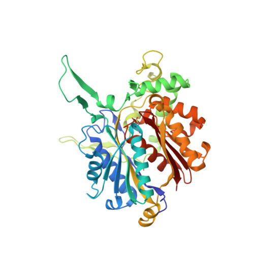
    Thiolases are CoA-dependent enzymes which catalyze the formation of a carbon-carbon bond in a Claisen condensation step and its reverse reaction via a thiolytic degradation mechanism. Mitochondrial acetoacetyl-coenzyme A (CoA) thiolase (T2) is important in the pathways for the synthesis and degradation of ketone bodies as well as for the degradation of 2-methylacetoacetyl-CoA. Human T2 deficiency has been identified in more than 60 patients. A unique property of T2 is its activation by potassium ions. High-resolution human T2 crystal structures are reported for the apo form and the CoA complex, with and without a bound potassium ion. The potassium ion is bound near the CoA binding site and the catalytic site. Binding of the potassium ion at this low-affinity binding site causes the rigidification of a CoA binding loop and an active site loop. Unexpectedly, a high-affinity binding site for a chloride ion has also been identified. The chloride ion is copurified, and its binding site is at the dimer interface, near two catalytic loops. A unique property of T2 is its ability to use 2-methyl-branched acetoacetyl-CoA as a substrate, whereas the other structurally characterized thiolases cannot utilize the 2-methylated compounds. The kinetic measurements show that T2 can degrade acetoacetyl-CoA and 2-methylacetoacetyl-CoA with similar catalytic efficiencies. For both substrates, the turnover numbers increase approximately 3-fold when the potassium ion concentration is increased from 0 to 40 mM KCl. The structural analysis of the active site of T2 indicates that the Phe325-Pro326 dipeptide near the catalytic cavity is responsible for the exclusive 2-methyl-branched substrate specificity.


  • Organizational Affiliation

    Biocenter Oulu and Department of Biochemistry, University of Oulu, P.O. Box 3000, FIN-90014 Oulu, Finland.


Macromolecules
Find similar proteins by:  (by identity cutoff)  |  3D Structure
Entity ID: 1
MoleculeChains Sequence LengthOrganismDetailsImage
Acetyl-CoA acetyltransferase
A, B, C, D
395Homo sapiensMutation(s): 2 
Gene Names: ACAT1
EC: 2.3.1.9
UniProt & NIH Common Fund Data Resources
Find proteins for P24752 (Homo sapiens)
Explore P24752 
Go to UniProtKB:  P24752
PHAROS:  P24752
GTEx:  ENSG00000075239 
Entity Groups  
Sequence Clusters30% Identity50% Identity70% Identity90% Identity95% Identity100% Identity
UniProt GroupP24752
Sequence Annotations
Expand
  • Reference Sequence
Small Molecules
Ligands 5 Unique
IDChains Name / Formula / InChI Key2D Diagram3D Interactions
COA
Query on COA

Download Ideal Coordinates CCD File 
G [auth A],
L [auth B],
P [auth C],
S [auth D]
COENZYME A
C21 H36 N7 O16 P3 S
RGJOEKWQDUBAIZ-IBOSZNHHSA-N
MES
Query on MES

Download Ideal Coordinates CCD File 
H [auth A]2-(N-MORPHOLINO)-ETHANESULFONIC ACID
C6 H13 N O4 S
SXGZJKUKBWWHRA-UHFFFAOYSA-N
GOL
Query on GOL

Download Ideal Coordinates CCD File 
I [auth A],
M [auth B]
GLYCEROL
C3 H8 O3
PEDCQBHIVMGVHV-UHFFFAOYSA-N
K
Query on K

Download Ideal Coordinates CCD File 
F [auth A],
K [auth B],
O [auth C],
R [auth D]
POTASSIUM ION
K
NPYPAHLBTDXSSS-UHFFFAOYSA-N
CL
Query on CL

Download Ideal Coordinates CCD File 
E [auth A],
J [auth B],
N [auth C],
Q [auth D]
CHLORIDE ION
Cl
VEXZGXHMUGYJMC-UHFFFAOYSA-M
Modified Residues  1 Unique
IDChains TypeFormula2D DiagramParent
CSO
Query on CSO
A, B, C, D
L-PEPTIDE LINKINGC3 H7 N O3 SCYS
Experimental Data & Validation

Experimental Data

  • Method: X-RAY DIFFRACTION
  • Resolution: 1.85 Å
  • R-Value Free: 0.203 
  • R-Value Work: 0.164 
  • R-Value Observed: 0.166 
  • Space Group: P 1 21 1
Unit Cell:
Length ( Å )Angle ( ˚ )
a = 75.349α = 90
b = 106.746β = 103.06
c = 101.66γ = 90
Software Package:
Software NamePurpose
REFMACrefinement
DNAdata collection
XDSdata reduction
XDSdata scaling
MOLREPphasing

Structure Validation

View Full Validation Report


Ligand Structure Quality Assessment 


Entry History 

Deposition Data

Revision History  (Full details and data files)

  • Version 1.0: 2007-04-03
    Type: Initial release
  • Version 1.1: 2008-05-01
    Changes: Version format compliance
  • Version 1.2: 2011-07-13
    Changes: Non-polymer description, Version format compliance
  • Version 1.3: 2021-11-10
    Changes: Database references, Derived calculations
  • Version 1.4: 2023-10-25
    Changes: Data collection, Refinement description
  • Version 1.5: 2023-11-15
    Changes: Data collection