2I88

Crystal structure of the Channel-forming Domain of Colicin E1


Experimental Data Snapshot

  • Method: X-RAY DIFFRACTION
  • Resolution: 2.50 Å
  • R-Value Free: 0.284 
  • R-Value Work: 0.172 
  • R-Value Observed: 0.176 

wwPDB Validation   3D Report Full Report


This is version 1.4 of the entry. See complete history


Literature

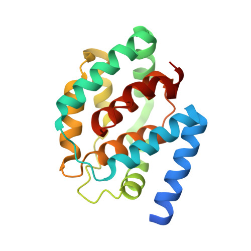
A mechanism for toxin insertion into membranes is suggested by the crystal structure of the channel-forming domain of colicin E1

Elkins, P.Bunker, A.Cramer, W.A.Stauffacher, C.V.

(1997) Structure 5: 443-458

  • DOI: https://doi.org/10.1016/s0969-2126(97)00200-1
  • Primary Citation of Related Structures:  
    2I88

  • PubMed Abstract: 

    Channel-forming colicins, including colicin E1, are a sub-family of bacteriocins. The toxic action of colicin E1 is derived from its ability to form a voltage-gated channel, which causes depolarization of the cytoplasmic membrane of sensitive Escherichia coli cells. In this process, the toxin-like colicin E1 molecule must undergo a substantial structural transition from a soluble state, in which it binds the target cell, to a membrane-bound state. Details of the structural changes that accompany this conversion may be directly applicable to other channel-forming toxins, as well as to the mechanism by which proteins insert into or cross membranes.


  • Organizational Affiliation

    Protein Engineering, Department Genentech, Inc. 460 Pt. San Bruno Blvd, South San Francisco, CA 94080, USA.


Macromolecules
Find similar proteins by:  (by identity cutoff)  |  3D Structure
Entity ID: 1
MoleculeChains Sequence LengthOrganismDetailsImage
Colicin-E1191Escherichia coliMutation(s): 0 
Gene Names: cea
Membrane Entity: Yes 
UniProt
Find proteins for P02978 (Escherichia coli)
Explore P02978 
Go to UniProtKB:  P02978
Entity Groups  
Sequence Clusters30% Identity50% Identity70% Identity90% Identity95% Identity100% Identity
UniProt GroupP02978
Sequence Annotations
Expand
  • Reference Sequence
Experimental Data & Validation

Experimental Data

  • Method: X-RAY DIFFRACTION
  • Resolution: 2.50 Å
  • R-Value Free: 0.284 
  • R-Value Work: 0.172 
  • R-Value Observed: 0.176 
  • Space Group: I 4
Unit Cell:
Length ( Å )Angle ( ˚ )
a = 87.46α = 90
b = 87.46β = 90
c = 59.1γ = 90
Software Package:
Software NamePurpose
CNSrefinement
PDB_EXTRACTdata extraction
CrystalCleardata collection
DENZOdata reduction
CCP4data scaling
CCP4phasing

Structure Validation

View Full Validation Report


Entry History 

Deposition Data

Revision History  (Full details and data files)

  • Version 1.0: 2006-09-26
    Type: Initial release
  • Version 1.1: 2008-05-01
    Changes: Version format compliance
  • Version 1.2: 2011-07-13
    Changes: Version format compliance
  • Version 1.3: 2018-01-24
    Changes: Advisory, Structure summary
  • Version 1.4: 2024-02-21
    Changes: Advisory, Data collection, Database references