2I3T

Bub3 complex with Mad3 (BubR1) GLEBS motif


Experimental Data Snapshot

  • Method: X-RAY DIFFRACTION
  • Resolution: 2.80 Å
  • R-Value Free: 0.267 
  • R-Value Work: 0.238 
  • R-Value Observed: 0.242 

wwPDB Validation   3D Report Full Report


This is version 1.3 of the entry. See complete history


Literature

Structural analysis of Bub3 interactions in the mitotic spindle checkpoint.

Larsen, N.A.Al-Bassam, J.Wei, R.R.Harrison, S.C.

(2007) Proc Natl Acad Sci U S A 104: 1201-1206

  • DOI: https://doi.org/10.1073/pnas.0610358104
  • Primary Citation of Related Structures:  
    2I3S, 2I3T

  • PubMed Abstract: 

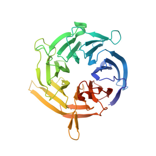
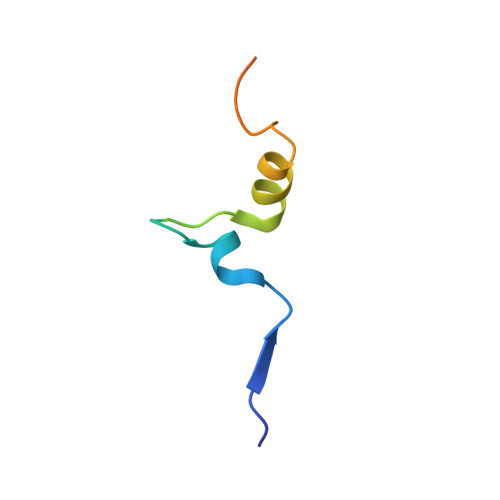
    The Mad3/BubR1, Mad2, Bub1, and Bub3 proteins are gatekeepers for the transition from metaphase to anaphase. Mad3 from Saccharomyces cerevisiae has homology to Bub1 but lacks a corresponding C-terminal kinase domain. Mad3 forms a stable heterodimer with Bub3. Negative-stain electron microscopy shows that Mad3 is an extended molecule (approximately 200 A long), whereas Bub3 is globular. The Gle2-binding-sequence (GLEBS) motifs found in Mad3 and Bub1 are necessary and sufficient for interaction with Bub3. The calorimetrically determined dissociation constants for GLEBS-motif peptides and Bub3 are approximately 5 microM. Crystal structures of these peptides with Bub3 show that the interactions for Mad3 and Bub1 are similar and mutually exclusive. In both structures, the GLEBS peptide snakes along the top surface of the beta-propeller, forming an extensive interface. Mutations in either protein that disrupt the interface cause checkpoint deficiency and chromosome instability. We propose that the structure imposed on the GLEBS segment by its association with Bub3 enables recruitment to unattached kinetochores.


  • Organizational Affiliation

    Jack Eileen Connors Structural Biology Laboratory, and Howard Hughes Medical Institute, Harvard Medical School, 250 Longwood Avenue, Boston, MA 02115, USA.


Macromolecules
Find similar proteins by:  (by identity cutoff)  |  3D Structure
Entity ID: 1
MoleculeChains Sequence LengthOrganismDetailsImage
Cell cycle arrest protein
A, C, E, G
341Saccharomyces cerevisiaeMutation(s): 0 
Gene Names: BUB3YOR026WOR26.16
UniProt
Find proteins for P26449 (Saccharomyces cerevisiae (strain ATCC 204508 / S288c))
Explore P26449 
Go to UniProtKB:  P26449
Entity Groups  
Sequence Clusters30% Identity50% Identity70% Identity90% Identity95% Identity100% Identity
UniProt GroupP26449
Sequence Annotations
Expand
  • Reference Sequence
Find similar proteins by:  (by identity cutoff)  |  3D Structure
Entity ID: 2
MoleculeChains Sequence LengthOrganismDetailsImage
Spindle assembly checkpoint component
B, D, F, H
54Saccharomyces cerevisiaeMutation(s): 0 
Gene Names: MAD3YJL013CJ1341
UniProt
Find proteins for P47074 (Saccharomyces cerevisiae (strain ATCC 204508 / S288c))
Explore P47074 
Go to UniProtKB:  P47074
Entity Groups  
Sequence Clusters30% Identity50% Identity70% Identity90% Identity95% Identity100% Identity
UniProt GroupP47074
Sequence Annotations
Expand
  • Reference Sequence
Experimental Data & Validation

Experimental Data

  • Method: X-RAY DIFFRACTION
  • Resolution: 2.80 Å
  • R-Value Free: 0.267 
  • R-Value Work: 0.238 
  • R-Value Observed: 0.242 
  • Space Group: P 1 21 1
Unit Cell:
Length ( Å )Angle ( ˚ )
a = 51.352α = 90
b = 173.335β = 95.3
c = 90.019γ = 90
Software Package:
Software NamePurpose
HKL-2000data collection
MOLREPphasing
CNSrefinement
HKL-2000data reduction
HKL-2000data scaling

Structure Validation

View Full Validation Report


Entry History 

Deposition Data

Revision History  (Full details and data files)

  • Version 1.0: 2007-01-09
    Type: Initial release
  • Version 1.1: 2008-05-01
    Changes: Version format compliance
  • Version 1.2: 2011-07-13
    Changes: Version format compliance
  • Version 1.3: 2023-08-30
    Changes: Data collection, Database references, Refinement description