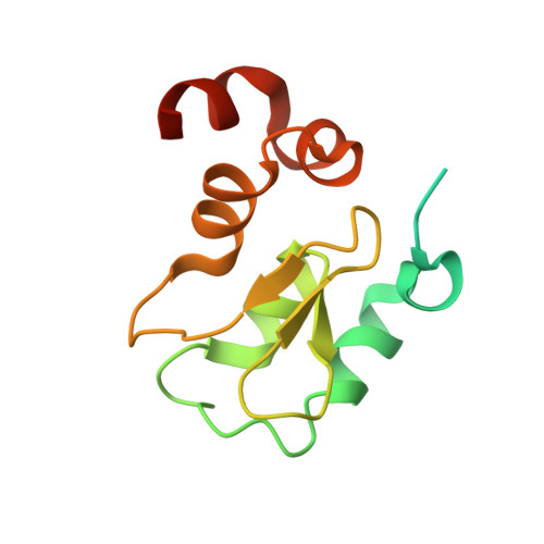
2I3I

Structure of an ML-IAP/XIAP chimera bound to a peptidomimetic


Experimental Data Snapshot

  • Method: X-RAY DIFFRACTION
  • Resolution: 2.30 Å
  • R-Value Free: 0.224 
  • R-Value Work: 0.188 
  • R-Value Observed: 0.190 

wwPDB Validation   3D Report Full Report


Ligand Structure Quality Assessment 


This is version 1.5 of the entry. See complete history


Literature

Design, synthesis, and biological activity of a potent Smac mimetic that sensitizes cancer cells to apoptosis by antagonizing IAPs.

Zobel, K.Wang, L.Varfolomeev, E.Franklin, M.C.Elliott, L.O.Wallweber, H.J.Okawa, D.C.Flygare, J.A.Vucic, D.Fairbrother, W.J.Deshayes, K.

(2006) ACS Chem Biol 1: 525-533

  • DOI: https://doi.org/10.1021/cb600276q
  • Primary Citation of Related Structures:  
    2I3H, 2I3I

  • PubMed Abstract: 

    Designed second mitochondrial activator of caspases (Smac) mimetics based on an accessible [7,5]-bicyclic scaffold bind to and antagonize protein interactions involving the inhibitor of apoptosis (IAP) proteins, X-chromosome-linked IAP (XIAP), melanoma IAP (ML-IAP), and c-IAPs 1 and 2 (cIAP1 and cIAP2). The design rationale is based on a combination of phage-panning data, peptide binding studies, and a survey of potential isosteres. The synthesis of two scaffolds is described. These compounds bind the XIAP-baculoviral IAP repeat 3 (BIR3), cIAP1-BIR3, cIAP2-BIR3, and ML-IAP-BIR domains with submicromolar affinities. The most potent Smac mimetic binds the cIAP1-BIR3 and ML-IAP-BIR domains with a K i of 50 nM. The X-ray crystal structure of this compound bound to an ML-IAP/XIAP chimeric BIR domain protein is compared with that of a complex with a phage-derived tetrapeptide, AVPW. The structures show that these compounds bind to the Smac-binding site on ML-IAP with identical hydrogen-bonding patterns and similar hydrophobic interactions. Consistent with the structural data, coimmunoprecipitation experiments demonstrate that the compounds can effectively block Smac interactions with ML-IAP. The compounds are further demonstrated to activate caspase-3 and -7, to reduce cell viability in assays using MDA-MB-231 breast cancer cells and A2058 melanoma cells, and to enhance doxorubicin-induced apoptosis in MDA-MB-231 cells.


  • Organizational Affiliation

    Department of Protein Engineering, Genentech, Inc., South San Francisco, California 94080, USA.


Macromolecules
Find similar proteins by:  (by identity cutoff)  |  3D Structure
Entity ID: 1
MoleculeChains Sequence LengthOrganismDetailsImage
Baculoviral IAP repeat-containing protein 7
A, B
133Homo sapiensMutation(s): 0 
Gene Names: BIRC7
UniProt & NIH Common Fund Data Resources
Find proteins for Q96CA5 (Homo sapiens)
Explore Q96CA5 
Go to UniProtKB:  Q96CA5
PHAROS:  Q96CA5
GTEx:  ENSG00000101197 
Entity Groups  
Sequence Clusters30% Identity50% Identity70% Identity90% Identity95% Identity100% Identity
UniProt GroupQ96CA5
Sequence Annotations
Expand
  • Reference Sequence
Small Molecules
Ligands 5 Unique
IDChains Name / Formula / InChI Key2D Diagram3D Interactions
618
Query on 618

Download Ideal Coordinates CCD File 
D [auth A],
G [auth B]
(3R,6R,9AR)-2,2-DIMETHYL-6-[(N-METHYL-L-ALANYL)AMINO]-N-(3-METHYL-1-PHENYL-1H-PYRAZOL-5-YL)-5-OXO-2,3,5,6,9,9A-HEXAHYDRO[1,3]THIAZOLO[3,2-A]AZEPINE-3-CARBOXAMIDE
C25 H32 N6 O3 S
WIYCWFZPNIUIGJ-RCVZYCBYSA-N
BTB
Query on BTB

Download Ideal Coordinates CCD File 
H [auth B]2-[BIS-(2-HYDROXY-ETHYL)-AMINO]-2-HYDROXYMETHYL-PROPANE-1,3-DIOL
C8 H19 N O5
OWMVSZAMULFTJU-UHFFFAOYSA-N
ZN
Query on ZN

Download Ideal Coordinates CCD File 
C [auth A],
E [auth B]
ZINC ION
Zn
PTFCDOFLOPIGGS-UHFFFAOYSA-N
EDO
Query on EDO

Download Ideal Coordinates CCD File 
I [auth B],
J [auth B]
1,2-ETHANEDIOL
C2 H6 O2
LYCAIKOWRPUZTN-UHFFFAOYSA-N
LI
Query on LI

Download Ideal Coordinates CCD File 
F [auth B]LITHIUM ION
Li
HBBGRARXTFLTSG-UHFFFAOYSA-N
Binding Affinity Annotations 
IDSourceBinding Affinity
618 PDBBind:  2I3I Ki: 50 (nM) from 1 assay(s)
Binding MOAD:  2I3I Ki: 50 (nM) from 1 assay(s)
Experimental Data & Validation

Experimental Data

  • Method: X-RAY DIFFRACTION
  • Resolution: 2.30 Å
  • R-Value Free: 0.224 
  • R-Value Work: 0.188 
  • R-Value Observed: 0.190 
  • Space Group: P 41 21 2
Unit Cell:
Length ( Å )Angle ( ˚ )
a = 87.356α = 90
b = 87.356β = 90
c = 73.196γ = 90
Software Package:
Software NamePurpose
REFMACrefinement
MAR345data collection
DENZOdata reduction
SCALEPACKdata scaling

Structure Validation

View Full Validation Report


Ligand Structure Quality Assessment 


Entry History 

Deposition Data

Revision History  (Full details and data files)

  • Version 1.0: 2006-09-19
    Type: Initial release
  • Version 1.1: 2008-05-01
    Changes: Version format compliance
  • Version 1.2: 2011-07-13
    Changes: Advisory, Version format compliance
  • Version 1.3: 2017-10-18
    Changes: Refinement description
  • Version 1.4: 2024-02-21
    Changes: Data collection, Database references, Derived calculations
  • Version 1.5: 2024-04-03
    Changes: Refinement description