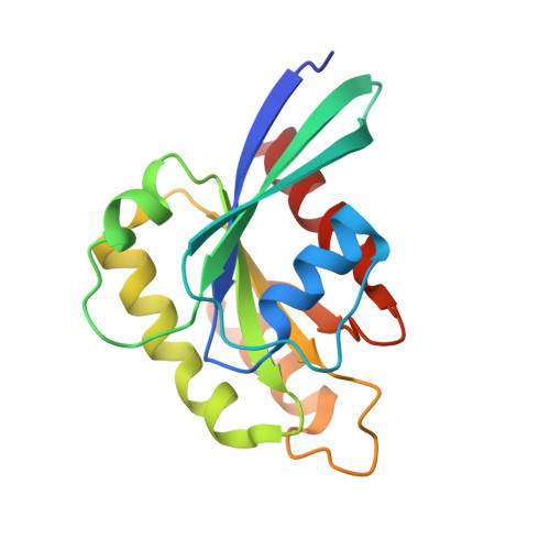
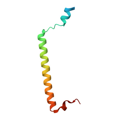
2HV8

Crystal structure of GTP-bound Rab11 in complex with FIP3


Experimental Data Snapshot

  • Method: X-RAY DIFFRACTION
  • Resolution: 1.86 Å
  • R-Value Free: 0.237 
  • R-Value Work: 0.203 
  • R-Value Observed: 0.205 

wwPDB Validation   3D Report Full Report


Ligand Structure Quality Assessment 


This is version 1.4 of the entry. See complete history


Literature

Structural Basis for Rab11-mediated Recruitment of FIP3 to Recycling Endosomes.

Eathiraj, S.Mishra, A.Prekeris, R.Lambright, D.G.

(2006) J Mol Biol 364: 121-135

  • DOI: https://doi.org/10.1016/j.jmb.2006.08.064
  • Primary Citation of Related Structures:  
    2HV8

  • PubMed Abstract: 

    The Rab11 GTPase regulates recycling of internalized plasma membrane receptors and is essential for completion of cytokinesis. A family of Rab11 interacting proteins (FIPs) that conserve a C-terminal Rab-binding domain (RBD) selectively recognize the active form of Rab11. Normal completion of cytokinesis requires a complex between Rab11 and FIP3. Here, we report the crystal structure and mutational analysis of a heterotetrameric complex between constitutively active Rab11 and a FIP3 construct that includes the RBD. Two Rab11 molecules bind to dyad symmetric sites at the C terminus of FIP3, which forms a non-canonical coiled-coiled dimer with a flared C terminus and hook region. The RBD overlaps with the coiled coil and extends through the C-terminal hook. Although FIP3 engages the switch and interswitch regions of Rab11, the mode of interaction differs significantly from that of other Rab-effector complexes. In particular, the switch II region undergoes a large structural rearrangement from an ordered but non-complementary active conformation to a remodeled conformation that facilitates the interaction with FIP3. Finally, we provide evidence that FIP3 can form homo-oligomers in cells, and that a critical determinant of Rab11 binding in vitro is necessary for FIP3 recruitment to recycling endosomes during cytokinesis.


  • Organizational Affiliation

    Program in Molecular Medicine and Department of Biochemistry & Molecular Pharmacology, University of Massachusetts Medical School, Worcester, MA 01605, USA.


Macromolecules
Find similar proteins by:  (by identity cutoff)  |  3D Structure
Entity ID: 1
MoleculeChains Sequence LengthOrganismDetailsImage
Ras-related protein Rab-11AA,
C [auth B],
E [auth C]
172Homo sapiensMutation(s): 1 
Gene Names: RAB11A
UniProt & NIH Common Fund Data Resources
Find proteins for P62491 (Homo sapiens)
Explore P62491 
Go to UniProtKB:  P62491
PHAROS:  P62491
GTEx:  ENSG00000103769 
Entity Groups  
Sequence Clusters30% Identity50% Identity70% Identity90% Identity95% Identity100% Identity
UniProt GroupP62491
Sequence Annotations
Expand
  • Reference Sequence
Find similar proteins by:  (by identity cutoff)  |  3D Structure
Entity ID: 2
MoleculeChains Sequence LengthOrganismDetailsImage
Rab11 family-interacting protein 3B [auth D],
D [auth E],
F
64Homo sapiensMutation(s): 0 
Gene Names: RAB11FIP3
UniProt & NIH Common Fund Data Resources
Find proteins for O75154 (Homo sapiens)
Explore O75154 
Go to UniProtKB:  O75154
PHAROS:  O75154
GTEx:  ENSG00000090565 
Entity Groups  
Sequence Clusters30% Identity50% Identity70% Identity90% Identity95% Identity100% Identity
UniProt GroupO75154
Sequence Annotations
Expand
  • Reference Sequence
Small Molecules
Ligands 5 Unique
IDChains Name / Formula / InChI Key2D Diagram3D Interactions
GTP
Query on GTP

Download Ideal Coordinates CCD File 
I [auth A],
N [auth B],
S [auth C]
GUANOSINE-5'-TRIPHOSPHATE
C10 H16 N5 O14 P3
XKMLYUALXHKNFT-UUOKFMHZSA-N
MES
Query on MES

Download Ideal Coordinates CCD File 
O [auth B]2-(N-MORPHOLINO)-ETHANESULFONIC ACID
C6 H13 N O4 S
SXGZJKUKBWWHRA-UHFFFAOYSA-N
SO4
Query on SO4

Download Ideal Coordinates CCD File 
H [auth A],
L [auth B],
M [auth B],
Q [auth C],
R [auth C]
SULFATE ION
O4 S
QAOWNCQODCNURD-UHFFFAOYSA-L
2ME
Query on 2ME

Download Ideal Coordinates CCD File 
J [auth B]METHOXYETHANE
C3 H8 O
XOBKSJJDNFUZPF-UHFFFAOYSA-N
MG
Query on MG

Download Ideal Coordinates CCD File 
G [auth A],
K [auth B],
P [auth C]
MAGNESIUM ION
Mg
JLVVSXFLKOJNIY-UHFFFAOYSA-N
Experimental Data & Validation

Experimental Data

  • Method: X-RAY DIFFRACTION
  • Resolution: 1.86 Å
  • R-Value Free: 0.237 
  • R-Value Work: 0.203 
  • R-Value Observed: 0.205 
  • Space Group: C 1 2 1
Unit Cell:
Length ( Å )Angle ( ˚ )
a = 218.291α = 90
b = 52.601β = 105.56
c = 70.679γ = 90
Software Package:
Software NamePurpose
REFMACrefinement
DENZOdata reduction
SCALEPACKdata scaling
AMoREphasing

Structure Validation

View Full Validation Report


Ligand Structure Quality Assessment 


Entry History 

Deposition Data

Revision History  (Full details and data files)

  • Version 1.0: 2006-11-21
    Type: Initial release
  • Version 1.1: 2008-05-01
    Changes: Version format compliance
  • Version 1.2: 2011-07-13
    Changes: Version format compliance
  • Version 1.3: 2021-10-20
    Changes: Database references, Derived calculations
  • Version 1.4: 2023-08-30
    Changes: Data collection, Refinement description