2HRC

1.7 angstrom structure of human ferrochelatase variant R115L


Experimental Data Snapshot

  • Method: X-RAY DIFFRACTION
  • Resolution: 1.70 Å
  • R-Value Free: 0.242 
  • R-Value Work: 0.221 
  • R-Value Observed: 0.221 

wwPDB Validation   3D Report Full Report


Ligand Structure Quality Assessment 


This is version 1.4 of the entry. See complete history


Literature

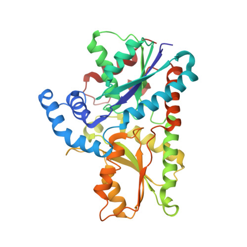
Substrate interactions with human ferrochelatase

Medlock, A.Swartz, L.Dailey, T.A.Dailey, H.A.Lanzilotta, W.N.

(2007) Proc Natl Acad Sci U S A 104: 1789-1793

  • DOI: https://doi.org/10.1073/pnas.0606144104
  • Primary Citation of Related Structures:  
    2HRC, 2HRE

  • PubMed Abstract: 

    Ferrochelatase, the terminal enzyme in heme biosynthesis, catalyzes the insertion of ferrous iron into protoporphyrin IX to form protoheme IX. Human ferrochelatase is a homodimeric, inner mitochondrial membrane-associated enzyme that possesses an essential [2Fe-2S] cluster. In this work, we report the crystal structure of human ferrochelatase with the substrate protoporphyrin IX bound as well as a higher resolution structure of the R115L variant without bound substrate. The data presented reveal that the porphyrin substrate is bound deep within an enclosed pocket. When compared with the location of N-methylmesoporphyrin in the Bacillus subtilis ferrochelatase, the porphyrin is rotated by approximately 100 degrees and is buried an additional 4.5 A deeper within the active site. The propionate groups of the substrate do not protrude into solvent and are bound in a manner similar to what has been observed in uroporphyrinogen decarboxylase. Furthermore, in the substrate-bound form, the jaws of the active site mouth are closed so that the porphyrin substrate is completely engulfed in the pocket. These data provide insights that will aid in the determination of the mechanism for ferrochelatase.


  • Organizational Affiliation

    Department of Biochemistry and Molecular Biology, University of Georgia, Athens, GA 30602, USA.


Macromolecules
Find similar proteins by:  (by identity cutoff)  |  3D Structure
Entity ID: 1
MoleculeChains Sequence LengthOrganismDetailsImage
Ferrochelatase
A, B
359Homo sapiensMutation(s): 1 
Gene Names: FECH
EC: 4.99.1.1
UniProt & NIH Common Fund Data Resources
Find proteins for P22830 (Homo sapiens)
Explore P22830 
Go to UniProtKB:  P22830
PHAROS:  P22830
GTEx:  ENSG00000066926 
Entity Groups  
Sequence Clusters30% Identity50% Identity70% Identity90% Identity95% Identity100% Identity
UniProt GroupP22830
Sequence Annotations
Expand
  • Reference Sequence
Small Molecules
Ligands 5 Unique
IDChains Name / Formula / InChI Key2D Diagram3D Interactions
CHD
Query on CHD

Download Ideal Coordinates CCD File 
F [auth A]
G [auth A]
H [auth A]
M [auth B]
N [auth B]
F [auth A],
G [auth A],
H [auth A],
M [auth B],
N [auth B],
O [auth B]
CHOLIC ACID
C24 H40 O5
BHQCQFFYRZLCQQ-OELDTZBJSA-N
FES
Query on FES

Download Ideal Coordinates CCD File 
D [auth A],
K [auth B]
FE2/S2 (INORGANIC) CLUSTER
Fe2 S2
NIXDOXVAJZFRNF-UHFFFAOYSA-N
GOL
Query on GOL

Download Ideal Coordinates CCD File 
I [auth A]GLYCEROL
C3 H8 O3
PEDCQBHIVMGVHV-UHFFFAOYSA-N
IMD
Query on IMD

Download Ideal Coordinates CCD File 
E [auth A],
L [auth B]
IMIDAZOLE
C3 H5 N2
RAXXELZNTBOGNW-UHFFFAOYSA-O
CL
Query on CL

Download Ideal Coordinates CCD File 
C [auth A],
J [auth B]
CHLORIDE ION
Cl
VEXZGXHMUGYJMC-UHFFFAOYSA-M
Experimental Data & Validation

Experimental Data

  • Method: X-RAY DIFFRACTION
  • Resolution: 1.70 Å
  • R-Value Free: 0.242 
  • R-Value Work: 0.221 
  • R-Value Observed: 0.221 
  • Space Group: P 21 21 21
Unit Cell:
Length ( Å )Angle ( ˚ )
a = 88.526α = 90
b = 92.983β = 90
c = 110.454γ = 90
Software Package:
Software NamePurpose
CNSrefinement
HKL-2000data reduction
HKL-2000data scaling
CNSphasing

Structure Validation

View Full Validation Report


Ligand Structure Quality Assessment 


Entry History 

Deposition Data

Revision History  (Full details and data files)

  • Version 1.0: 2007-03-13
    Type: Initial release
  • Version 1.1: 2008-05-01
    Changes: Version format compliance
  • Version 1.2: 2011-07-13
    Changes: Non-polymer description, Version format compliance
  • Version 1.3: 2021-10-20
    Changes: Database references, Derived calculations
  • Version 1.4: 2023-08-30
    Changes: Data collection, Refinement description