2HD6

Crystal structure of the human carbonic anhydrase II in complex with a hypoxia-activatable sulfonamide.


Experimental Data Snapshot

  • Method: X-RAY DIFFRACTION
  • Resolution: 1.80 Å
  • R-Value Free: 0.217 
  • R-Value Work: 0.184 

wwPDB Validation   3D Report Full Report


Ligand Structure Quality Assessment 


This is version 1.3 of the entry. See complete history


Literature

Carbonic anhydrase inhibitors: Hypoxia-activatable sulfonamides incorporating disulfide bonds that target the tumor-associated isoform IX.

De Simone, G.Vitale, R.M.Di Fiore, A.Pedone, C.Scozzafava, A.Montero, J.L.Winum, J.Y.Supuran, C.T.

(2006) J Med Chem 49: 5544-5551

  • DOI: https://doi.org/10.1021/jm060531j
  • Primary Citation of Related Structures:  
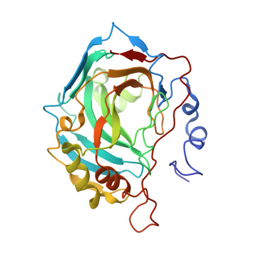
    2HD6

  • PubMed Abstract: 

    An approach for designing bioreductive, hypoxia-activatable carbonic anhydrase (CA, EC 4.2.1.1) inhibitors targeting the tumor-associated isoforms is reported. Sulfonamides incorporating 3,3'-dithiodipropionamide/2,2'-dithiodibenzamido moieties were prepared and reduced enzymatically/chemically in conditions present in hypoxic tumors, leading to thiols. The X-ray crystal structure of the most promising compound, 4-(2-mercaptophenylcarboxamido)benzenesulfonamide, which as disulfide showed a K(I) against hCA IX of 653 nM (in reduced form of 9.1 nM), in adduct with hCA II showed the inhibitor making favorable interactions with Gln92, Val121, Phe131, Leu198, Thr199, Thr200, Pro201, and Pro202, whereas the sulfamoyl moiety was coordinated to the Zn2+ ion. The same interactions were preserved in the adduct with hCA IX, but in addition, a hydrogen bond between the SH moiety of the inhibitor and the amide nitrogen of Gln67 was evidenced, which may explain the almost 2 times more effective inhibition of the tumor-associated isozyme over the cytosolic isoform.


  • Organizational Affiliation

    Istituto di Biostrutture e Bioimmagini-CNR, Via Mezzocannone 16, 80134 Naples, Italy. gdesimone@unina.it


Macromolecules
Find similar proteins by:  (by identity cutoff)  |  3D Structure
Entity ID: 1
MoleculeChains Sequence LengthOrganismDetailsImage
Carbonic anhydrase 2259Homo sapiensMutation(s): 0 
EC: 4.2.1.1
UniProt & NIH Common Fund Data Resources
Find proteins for P00918 (Homo sapiens)
Explore P00918 
Go to UniProtKB:  P00918
PHAROS:  P00918
GTEx:  ENSG00000104267 
Entity Groups  
Sequence Clusters30% Identity50% Identity70% Identity90% Identity95% Identity100% Identity
UniProt GroupP00918
Sequence Annotations
Expand
  • Reference Sequence
Small Molecules
Ligands 5 Unique
IDChains Name / Formula / InChI Key2D Diagram3D Interactions
MBO
Query on MBO

Download Ideal Coordinates CCD File 
D [auth A]MERCURIBENZOIC ACID
C7 H5 Hg O2
FVFZSVRSDNUCGG-UHFFFAOYSA-N
BOS
Query on BOS

Download Ideal Coordinates CCD File 
E [auth A]N-[4-(AMINOSULFONYL)PHENYL]-2-MERCAPTOBENZAMIDE
C13 H12 N2 O3 S2
MRLVIVXGUGNENL-UHFFFAOYSA-N
GOL
Query on GOL

Download Ideal Coordinates CCD File 
F [auth A]GLYCEROL
C3 H8 O3
PEDCQBHIVMGVHV-UHFFFAOYSA-N
ZN
Query on ZN

Download Ideal Coordinates CCD File 
B [auth A]ZINC ION
Zn
PTFCDOFLOPIGGS-UHFFFAOYSA-N
CL
Query on CL

Download Ideal Coordinates CCD File 
C [auth A]CHLORIDE ION
Cl
VEXZGXHMUGYJMC-UHFFFAOYSA-M
Binding Affinity Annotations 
IDSourceBinding Affinity
BOS PDBBind:  2HD6 Ki: 16 (nM) from 1 assay(s)
Binding MOAD:  2HD6 Ki: 16 (nM) from 1 assay(s)
BindingDB:  2HD6 Ki: 16 (nM) from 1 assay(s)
CL BindingDB:  2HD6 Ki: min: 2.00e+8, max: 2.00e+8 (nM) from 2 assay(s)
Experimental Data & Validation

Experimental Data

  • Method: X-RAY DIFFRACTION
  • Resolution: 1.80 Å
  • R-Value Free: 0.217 
  • R-Value Work: 0.184 
  • Space Group: P 1 21 1
Unit Cell:
Length ( Å )Angle ( ˚ )
a = 42.07α = 90
b = 41.26β = 104.28
c = 71.66γ = 90
Software Package:
Software NamePurpose
CNSrefinement
CNSphasing

Structure Validation

View Full Validation Report


Ligand Structure Quality Assessment 


Entry History 

Deposition Data

Revision History  (Full details and data files)

  • Version 1.0: 2006-10-03
    Type: Initial release
  • Version 1.1: 2008-05-01
    Changes: Version format compliance
  • Version 1.2: 2011-07-13
    Changes: Non-polymer description, Version format compliance
  • Version 1.3: 2023-08-30
    Changes: Data collection, Database references, Derived calculations, Refinement description