2HB3

Wild-type HIV-1 Protease in complex with potent inhibitor GRL06579


Experimental Data Snapshot

  • Method: X-RAY DIFFRACTION
  • Resolution: 1.35 Å
  • R-Value Free: 0.192 
  • R-Value Work: 0.147 
  • R-Value Observed: 0.150 

wwPDB Validation   3D Report Full Report


Ligand Structure Quality Assessment 


This is version 1.4 of the entry. See complete history


Literature

Structure-Based Design of Novel HIV-1 Protease Inhibitors To Combat Drug Resistance.

Ghosh, A.K.Sridhar, P.R.Leshchenko, S.Hussain, A.K.Li, J.Kovalevsky, A.Y.Walters, D.E.Wedekind, J.E.Grum-Tokars, V.Das, D.Koh, Y.Maeda, K.Gatanaga, H.Weber, I.T.Mitsuya, H.

(2006) J Med Chem 49: 5252-5261

  • DOI: https://doi.org/10.1021/jm060561m
  • Primary Citation of Related Structures:  
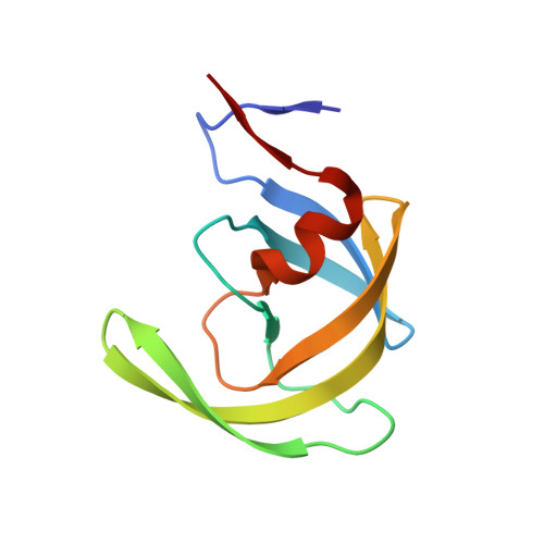
    2HB3

  • PubMed Abstract: 

    Structure-based design and synthesis of novel HIV protease inhibitors are described. The inhibitors are designed specifically to interact with the backbone of HIV protease active site to combat drug resistance. Inhibitor 3 has exhibited exceedingly potent enzyme inhibitory and antiviral potency. Furthermore, this inhibitor maintains impressive potency against a wide spectrum of HIV including a variety of multi-PI-resistant clinical strains. The inhibitors incorporated a stereochemically defined 5-hexahydrocyclopenta[b]furanyl urethane as the P2-ligand into the (R)-(hydroxyethylamino)sulfonamide isostere. Optically active (3aS,5R,6aR)-5-hydroxy-hexahydrocyclopenta[b]furan was prepared by an enzymatic asymmetrization of meso-diacetate with acetyl cholinesterase, radical cyclization, and Lewis acid-catalyzed anomeric reduction as the key steps. A protein-ligand X-ray crystal structure of inhibitor 3-bound HIV-1 protease (1.35 A resolution) revealed extensive interactions in the HIV protease active site including strong hydrogen bonding interactions with the backbone. This design strategy may lead to novel inhibitors that can combat drug resistance.


  • Organizational Affiliation

    Department of Chemistry and Medicinal Chemistry, Purdue University, West Lafayette, Indiana 47907, USA. akghosh@purdue.edu


Macromolecules
Find similar proteins by:  (by identity cutoff)  |  3D Structure
Entity ID: 1
MoleculeChains Sequence LengthOrganismDetailsImage
Protease
A, B
99Human immunodeficiency virus 1Mutation(s): 5 
Gene Names: GAGPOL
EC: 3.4.23.4
UniProt
Find proteins for Q7SSI0 (Human immunodeficiency virus 1)
Explore Q7SSI0 
Go to UniProtKB:  Q7SSI0
Entity Groups  
Sequence Clusters30% Identity50% Identity70% Identity90% Identity95% Identity100% Identity
UniProt GroupQ7SSI0
Sequence Annotations
Expand
  • Reference Sequence
Small Molecules
Ligands 4 Unique
IDChains Name / Formula / InChI Key2D Diagram3D Interactions
GRL
Query on GRL

Download Ideal Coordinates CCD File 
H [auth B](3AS,5R,6AR)-HEXAHYDRO-2H-CYCLOPENTA[B]FURAN-5-YL (2S,3S)-3-HYDROXY-4-(4-(HYDROXYMETHYL)-N-ISOBUTYLPHENYLSULFONAMIDO)-1-PHENYLBUTAN-2-YLCARBAMATE
C29 H40 N2 O7 S
VYBDPVQMILRSMK-GRXYLYAXSA-N
GOL
Query on GOL

Download Ideal Coordinates CCD File 
I [auth B]GLYCEROL
C3 H8 O3
PEDCQBHIVMGVHV-UHFFFAOYSA-N
CL
Query on CL

Download Ideal Coordinates CCD File 
C [auth A],
E [auth B],
F [auth B]
CHLORIDE ION
Cl
VEXZGXHMUGYJMC-UHFFFAOYSA-M
NA
Query on NA

Download Ideal Coordinates CCD File 
D [auth A],
G [auth B]
SODIUM ION
Na
FKNQFGJONOIPTF-UHFFFAOYSA-N
Binding Affinity Annotations 
IDSourceBinding Affinity
GRL PDBBind:  2HB3 Ki: 4.50e-3 (nM) from 1 assay(s)
BindingDB:  2HB3 Ki: min: 4.50e-3, max: 4.5 (nM) from 3 assay(s)
IC50: 1.8 (nM) from 1 assay(s)
Binding MOAD:  2HB3 Ki: 4.50e-3 (nM) from 1 assay(s)
Experimental Data & Validation

Experimental Data

  • Method: X-RAY DIFFRACTION
  • Resolution: 1.35 Å
  • R-Value Free: 0.192 
  • R-Value Work: 0.147 
  • R-Value Observed: 0.150 
  • Space Group: P 21 21 2
Unit Cell:
Length ( Å )Angle ( ˚ )
a = 58.08α = 90
b = 86.48β = 90
c = 45.94γ = 90
Software Package:
Software NamePurpose
SHELXmodel building
SHELXL-97refinement
ADSCdata collection
DENZOdata reduction
SCALEPACKdata scaling
AMoREphasing

Structure Validation

View Full Validation Report


Ligand Structure Quality Assessment 


Entry History 

Deposition Data

Revision History  (Full details and data files)

  • Version 1.0: 2006-08-29
    Type: Initial release
  • Version 1.1: 2008-05-01
    Changes: Version format compliance
  • Version 1.2: 2011-07-13
    Changes: Non-polymer description, Version format compliance
  • Version 1.3: 2021-10-20
    Changes: Database references, Derived calculations
  • Version 1.4: 2024-02-14
    Changes: Data collection