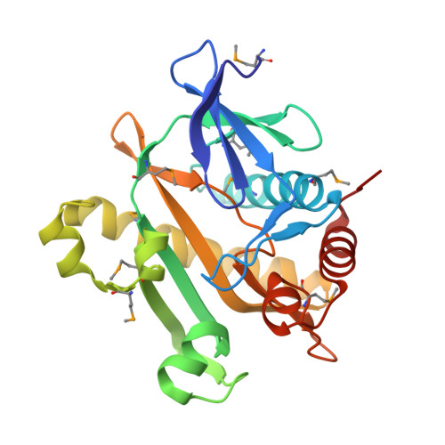
Nucleotide Complexes of Escherichia coli Phosphoribosylaminoimidazole Succinocarboxamide Synthetase.
Ginder, N.D., Binkowski, D.J., Fromm, H.J., Honzatko, R.B.(2006) J Biol Chem 281: 20680-20688
- PubMed: 16687397
- DOI: https://doi.org/10.1074/jbc.M602109200
- Primary Citation of Related Structures:
2GQR, 2GQS - PubMed Abstract:
Phosphoribosylaminoimidazole-succinocarboxamide synthetase (SAICAR synthetase) converts 4-carboxy-5-aminoimidazole ribonucleotide (CAIR) to 4-(N-succinylcarboxamide)-5-aminoimidazole ribonucleotide (SAICAR). The enzyme is a target of natural products that impair cell growth. Reported here are the crystal structures of the ADP and the ADP.CAIR complexes of SAICAR synthetase from Escherichia coli, the latter being the first instance of a CAIR-ligated SAICAR synthetase. ADP and CAIR bind to the active site in association with three Mg(2+), two of which coordinate the same oxygen atom of the 4-carboxyl group of CAIR; whereas, the third coordinates the alpha- and beta-phosphoryl groups of ADP. The ADP.CAIR complex is the basis for a transition state model of a phosphoryl transfer reaction involving CAIR and ATP, but also supports an alternative chemical pathway in which the nucleophilic attack of l-aspartate precedes the phosphoryl transfer reaction. The polypeptide fold for residues 204-221 of the E. coli structure differs significantly from those of the ligand-free SAICAR synthetase from Thermatoga maritima and the adenine nucleotide complexes of the synthetase from Saccharomyces cerevisiae. Conformational differences between the E. coli, T. maritima, and yeast synthetases suggest the possibility of selective inhibition of de novo purine nucleotide biosynthesis in microbial organisms.
Organizational Affiliation:
Department of Biochemistry, Biophysics, and Molecular Biology, Iowa State University, Ames, Iowa 50011.