2GKD

Structural insight into self-sacrifice mechanism of enediyne resistance


Experimental Data Snapshot

  • Method: SOLUTION NMR
  • Conformers Calculated: 100 
  • Conformers Submitted: 
  • Selection Criteria: structures with the least restraint violations 

wwPDB Validation   3D Report Full Report

Currently 2GKD does not have a validation slider image.


This is version 1.3 of the entry. See complete history


Literature

Structural insight into the self-sacrifice mechanism of enediyne resistance.

Singh, S.Hager, M.H.Zhang, C.Griffith, B.R.Lee, M.S.Hallenga, K.Markley, J.L.Thorson, J.S.

(2006) ACS Chem Biol 1: 451-460

  • DOI: https://doi.org/10.1021/cb6002898
  • Primary Citation of Related Structures:  
    1ZXF, 2GKD

  • PubMed Abstract: 

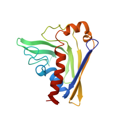
    The recent discovery of the first "self-sacrifice" mechanism for bacterial resistance to the enediyne antitumor antibiotics, where enediyne-induced proteolysis of the resistance protein CalC inactivates both the highly reactive metabolite and the resistance protein, revealed yet another ingenious bacterial mechanism for controlling reactive metabolites. As reported herein, the first 3D structures of CalC and CalC in complex with calicheamicin (CLM) divulge CalC to be a member of the steroidogenic acute regulatory protein (StAR)-related transfer (START) domain superfamily. In contrast to previous studies of proteins known to bind DNA-damaging natural products ( e.g ., bleomycins, mitomycins, and nine-membered chromoprotein enediynes), this is the first demonstrated involvement of a START domain fold. Consistent with the CalC self-sacrifice mechanism, CLM in complex with CalC is positioned for direct hydrogen abstraction from Gly113 to initiate the oxidative proteolysis-based resistance mechanism. These structural studies also illuminate, for the first time, a small DNA-binding region within CalC that may serve to localize CalC to the enediyne target (DNA). Given the role of START domains in nuclear/cytosolic transport and translocation, this structural study also may implicate START domains as post-endocytotic intracellular chaperones for enediyne-based therapeutics such as MyloTarg.


  • Organizational Affiliation

    Center for Eukaryotic Structural Genomics, Department of Biochemistry, University of Wisconsin-Madison, Madison, Wisconsin 53706-1544, USA.


Macromolecules

Find similar proteins by:  (by identity cutoff)  |  3D Structure
Entity ID: 3
MoleculeChains Sequence LengthOrganismDetailsImage
CalCC [auth A]155Micromonospora echinosporaMutation(s): 0 
UniProt
Find proteins for Q8KNF0 (Micromonospora echinospora)
Explore Q8KNF0 
Go to UniProtKB:  Q8KNF0
Entity Groups  
Sequence Clusters30% Identity50% Identity70% Identity90% Identity95% Identity100% Identity
UniProt GroupQ8KNF0
Sequence Annotations
Expand
  • Reference Sequence

Find similar nucleic acids by:  Sequence   |   3D Structure  

Entity ID: 1
MoleculeChains LengthOrganismImage
5'-D(*GP*CP*AP*TP*AP*TP*GP*AP*TP*AP*G)-3'A [auth B]11N/A
Sequence Annotations
Expand
  • Reference Sequence

Find similar nucleic acids by:  Sequence   |   3D Structure  

Entity ID: 2
MoleculeChains LengthOrganismImage
5'-D(*CP*TP*AP*TP*CP*AP*TP*AP*TP*GP*C)-3'B [auth C]11N/A
Sequence Annotations
Expand
  • Reference Sequence
Small Molecules
Binding Affinity Annotations 
IDSourceBinding Affinity
DNA PDBBind:  2GKD Kd: 1.40e+4 (nM) from 1 assay(s)
Experimental Data & Validation

Experimental Data

  • Method: SOLUTION NMR
  • Conformers Calculated: 100 
  • Conformers Submitted: 
  • Selection Criteria: structures with the least restraint violations 

Structure Validation

View Full Validation Report

Currently 2GKD does not have a validation slider image.


Entry History 

Deposition Data

Revision History  (Full details and data files)

  • Version 1.0: 2006-08-22
    Type: Initial release
  • Version 1.1: 2008-05-01
    Changes: Version format compliance
  • Version 1.2: 2011-07-13
    Changes: Version format compliance
  • Version 1.3: 2022-03-09
    Changes: Data collection, Database references, Derived calculations