2GGM

Human centrin 2 xeroderma pigmentosum group C protein complex


Experimental Data Snapshot

  • Method: X-RAY DIFFRACTION
  • Resolution: 2.35 Å
  • R-Value Free: 0.245 
  • R-Value Work: 0.192 
  • R-Value Observed: 0.194 

wwPDB Validation   3D Report Full Report


This is version 1.2 of the entry. See complete history


Literature

The structure of the human centrin 2-xeroderma pigmentosum group C protein complex.

Thompson, J.R.Ryan, Z.C.Salisbury, J.L.Kumar, R.

(2006) J Biol Chem 281: 18746-18752

  • DOI: https://doi.org/10.1074/jbc.M513667200
  • Primary Citation of Related Structures:  
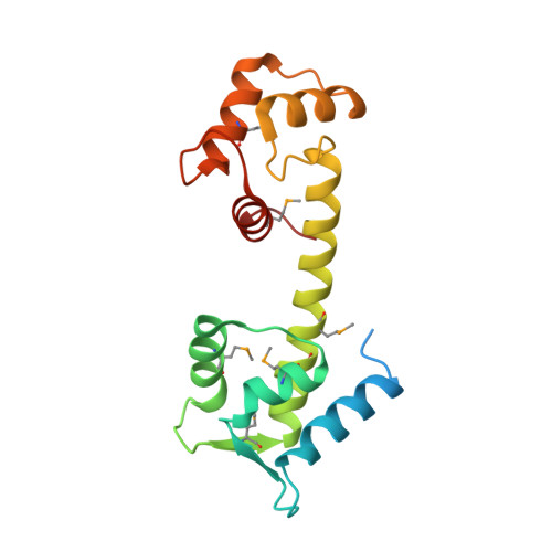
    2GGM

  • PubMed Abstract: 

    Human centrin-2 plays a key role in centrosome function and stimulates nucleotide excision repair by binding to the xeroderma pigmentosum group C protein. To determine the structure of human centrin-2 and to develop an understanding of molecular interactions between centrin and xeroderma pigmentosum group C protein, we characterized the crystal structure of calcium-loaded full-length centrin-2 complexed with a xeroderma pigmentosum group C peptide. Our structure shows that the carboxyl-terminal domain of centrin-2 binds this peptide and two calcium atoms, whereas the amino-terminal lobe is in a closed conformation positioned distantly by an ordered alpha-helical linker. A stretch of the amino-terminal domain unique to centrins appears disordered. Two xeroderma pigmentosum group C peptides both bound to centrin-2 also interact to form an alpha-helical coiled-coil. The interface between centrin-2 and each peptide is predominantly nonpolar, and key hydrophobic residues of XPC have been identified that lead us to propose a novel binding motif for centrin.


  • Organizational Affiliation

    Department of Physiology, Mayo Clinic College of Medicine, Rochester, Minnesota 55905, USA. thompson.james@mayo.edu


Macromolecules
Find similar proteins by:  (by identity cutoff)  |  3D Structure
Entity ID: 1
MoleculeChains Sequence LengthOrganismDetailsImage
Centrin-2
A, B
172Homo sapiensMutation(s): 7 
Gene Names: CETN2CALTCEN2
UniProt & NIH Common Fund Data Resources
Find proteins for P41208 (Homo sapiens)
Explore P41208 
Go to UniProtKB:  P41208
PHAROS:  P41208
GTEx:  ENSG00000147400 
Entity Groups  
Sequence Clusters30% Identity50% Identity70% Identity90% Identity95% Identity100% Identity
UniProt GroupP41208
Sequence Annotations
Expand
  • Reference Sequence

Find similar proteins by:  Sequence   |   3D Structure  

Entity ID: 2
MoleculeChains Sequence LengthOrganismDetailsImage
DNA-repair protein complementing XP-C cells
C, D
17N/AMutation(s): 0 
UniProt & NIH Common Fund Data Resources
Find proteins for Q01831 (Homo sapiens)
Explore Q01831 
Go to UniProtKB:  Q01831
PHAROS:  Q01831
GTEx:  ENSG00000154767 
Entity Groups  
Sequence Clusters30% Identity50% Identity70% Identity90% Identity95% Identity100% Identity
UniProt GroupQ01831
Sequence Annotations
Expand
  • Reference Sequence
Experimental Data & Validation

Experimental Data

  • Method: X-RAY DIFFRACTION
  • Resolution: 2.35 Å
  • R-Value Free: 0.245 
  • R-Value Work: 0.192 
  • R-Value Observed: 0.194 
  • Space Group: C 1 2 1
Unit Cell:
Length ( Å )Angle ( ˚ )
a = 59.15α = 90
b = 59β = 94.3
c = 104.24γ = 90
Software Package:
Software NamePurpose
REFMACrefinement
CBASSdata collection
HKL-2000data scaling
SOLVEphasing

Structure Validation

View Full Validation Report


Entry History 

Deposition Data

Revision History  (Full details and data files)

  • Version 1.0: 2006-04-25
    Type: Initial release
  • Version 1.1: 2008-05-01
    Changes: Version format compliance
  • Version 1.2: 2011-07-13
    Changes: Version format compliance