2G0C

Structure of the RNA binding domain (residues 404-479) of the Bacillus subtilis YxiN protein


Experimental Data Snapshot

  • Method: X-RAY DIFFRACTION
  • Resolution: 1.70 Å
  • R-Value Free: 0.276 
  • R-Value Work: 0.253 
  • R-Value Observed: 0.253 

wwPDB Validation   3D Report Full Report


This is version 1.5 of the entry. See complete history


Literature

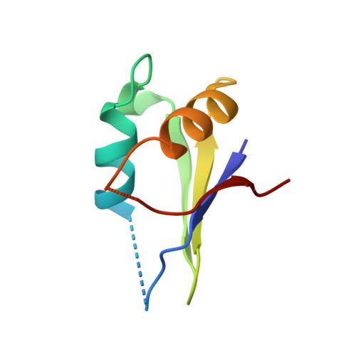
The domain of the Bacillus subtilis DEAD-box helicase YxiN that is responsible for specific binding of 23S rRNA has an RNA recognition motif fold.

Wang, S.Hu, Y.Overgaard, M.T.Karginov, F.V.Uhlenbeck, O.C.McKay, D.B.

(2006) RNA 12: 959-967

  • DOI: https://doi.org/10.1261/rna.5906
  • Primary Citation of Related Structures:  
    2G0C

  • PubMed Abstract: 

    The YxiN protein of Bacillus subtilis is a member of the DbpA subfamily of prokaryotic DEAD-box RNA helicases. Like DbpA, it binds with high affinity and specificity to segments of 23S ribosomal RNA as short as 32 nucleotides (nt) that include hairpin 92. Several experiments have shown that the 76-residue carboxy-terminal domain of YxiN is responsible for the high-affinity RNA binding. The domain has been crystallized and its structure has been solved to 1.7 Angstroms resolution. The structure reveals an RNA recognition motif (RRM) fold that is found in many eukaryotic RNA binding proteins; the RRM fold was not apparent from the amino acid sequence. The domain has two solvent exposed aromatic residues at sites that correspond to the aromatic residues of the ribonucleoprotein (RNP) motifs RNP1 and RNP2 that are essential for RNA binding in many RRMs. However, mutagenesis of these residues (Tyr404 and Tyr447) to alanine has little effect on RNA affinity, suggesting that the YxiN domain binds target RNAs in a manner that differs from the binding mode commonly found in many eukaryotic RRMs.


  • Organizational Affiliation

    Department of Structural Biology, Stanford University School of Medicine, California 94305, USA.


Macromolecules
Find similar proteins by:  (by identity cutoff)  |  3D Structure
Entity ID: 1
MoleculeChains Sequence LengthOrganismDetailsImage
ATP-dependent RNA helicase dbpA76Bacillus subtilisMutation(s): 0 
Gene Names: dbpAdeaD
EC: 3.6.1
UniProt
Find proteins for P42305 (Bacillus subtilis (strain 168))
Explore P42305 
Go to UniProtKB:  P42305
Entity Groups  
Sequence Clusters30% Identity50% Identity70% Identity90% Identity95% Identity100% Identity
UniProt GroupP42305
Sequence Annotations
Expand
  • Reference Sequence
Experimental Data & Validation

Experimental Data

  • Method: X-RAY DIFFRACTION
  • Resolution: 1.70 Å
  • R-Value Free: 0.276 
  • R-Value Work: 0.253 
  • R-Value Observed: 0.253 
  • Space Group: P 41 21 2
Unit Cell:
Length ( Å )Angle ( ˚ )
a = 51.01α = 90
b = 51.01β = 90
c = 64.26γ = 90
Software Package:
Software NamePurpose
DENZOdata reduction
SCALEPACKdata scaling
CNSrefinement
PDB_EXTRACTdata extraction
HKL-2000data reduction
SOLVEphasing

Structure Validation

View Full Validation Report


Entry History 

Deposition Data

  • Released Date: 2006-04-18 
  • Deposition Author(s): McKay, D.B.

Revision History  (Full details and data files)

  • Version 1.0: 2006-04-18
    Type: Initial release
  • Version 1.1: 2008-05-01
    Changes: Version format compliance
  • Version 1.2: 2011-07-13
    Changes: Derived calculations, Version format compliance
  • Version 1.3: 2017-10-18
    Changes: Refinement description
  • Version 1.4: 2024-02-14
    Changes: Data collection, Database references, Derived calculations
  • Version 1.5: 2024-04-03
    Changes: Refinement description