2FW4

Carbonic anhydrase activators. The first X-ray crystallographic study of an activator of isoform I, structure with L-histidine.


Experimental Data Snapshot

  • Method: X-RAY DIFFRACTION
  • Resolution: 2.00 Å
  • R-Value Free: 0.303 
  • R-Value Work: 0.213 
  • R-Value Observed: 0.217 

wwPDB Validation   3D Report Full Report


Ligand Structure Quality Assessment 


This is version 1.4 of the entry. See complete history


Literature

Carbonic anhydrase activators: The first X-ray crystallographic study of an adduct of isoform I.

Temperini, C.Scozzafava, A.Supuran, C.T.

(2006) Bioorg Med Chem Lett 16: 5152-5156

  • DOI: https://doi.org/10.1016/j.bmcl.2006.07.021
  • Primary Citation of Related Structures:  
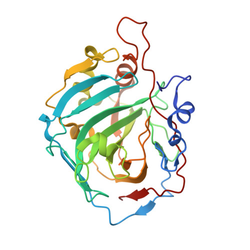
    2FW4

  • PubMed Abstract: 

    The X-ray crystallographic structure for the adduct of an activator with human carbonic anhydrase isozyme I (hCA I) is reported. L-Histidine binds deep within the enzyme active site, participating in a network of hydrogen bonds involving its carboxylate moiety and the zinc-bound water molecule, as well as the imidazole of His200, being in van der Waals contacts with Thr199, His200, His64, and His67. This binding is very different from that to the other major cytosolic isozyme hCA II.


  • Organizational Affiliation

    Università degli Studi di Firenze, Laboratorio di Chimica Bioinorganica, Rm. 188, Via della Lastruccia 3, I-50019 Sesto Fiorentino, Firenze, Italy.


Macromolecules
Find similar proteins by:  (by identity cutoff)  |  3D Structure
Entity ID: 1
MoleculeChains Sequence LengthOrganismDetailsImage
Carbonic anhydrase 1
A, B
260Homo sapiensMutation(s): 0 
EC: 4.2.1.1
UniProt & NIH Common Fund Data Resources
Find proteins for P00915 (Homo sapiens)
Explore P00915 
Go to UniProtKB:  P00915
PHAROS:  P00915
GTEx:  ENSG00000133742 
Entity Groups  
Sequence Clusters30% Identity50% Identity70% Identity90% Identity95% Identity100% Identity
UniProt GroupP00915
Sequence Annotations
Expand
  • Reference Sequence
Experimental Data & Validation

Experimental Data

  • Method: X-RAY DIFFRACTION
  • Resolution: 2.00 Å
  • R-Value Free: 0.303 
  • R-Value Work: 0.213 
  • R-Value Observed: 0.217 
  • Space Group: P 21 21 21
Unit Cell:
Length ( Å )Angle ( ˚ )
a = 62.138α = 90
b = 70.182β = 90
c = 120.573γ = 90
Software Package:
Software NamePurpose
REFMACrefinement
MOSFLMdata reduction
SCALEPACKdata scaling
AMoREphasing

Structure Validation

View Full Validation Report


Ligand Structure Quality Assessment 


Entry History 

Deposition Data

Revision History  (Full details and data files)

  • Version 1.0: 2006-08-08
    Type: Initial release
  • Version 1.1: 2008-05-01
    Changes: Version format compliance
  • Version 1.2: 2011-07-13
    Changes: Version format compliance
  • Version 1.3: 2018-04-04
    Changes: Data collection
  • Version 1.4: 2024-02-14
    Changes: Data collection, Database references, Derived calculations, Refinement description