The RNA binding domain of ribosomal protein L11: three-dimensional structure of the RNA-bound form of the protein and its interaction with 23 S rRNA.
Hinck, A.P., Markus, M.A., Huang, S., Grzesiek, S., Kustonovich, I., Draper, D.E., Torchia, D.A.(1997) J Mol Biol 274: 101-113
- PubMed: 9398519
- DOI: https://doi.org/10.1006/jmbi.1997.1379
- Primary Citation of Related Structures:
1FOY, 2FOW - PubMed Abstract:
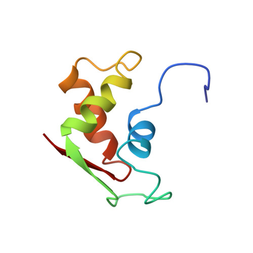
The three-dimensional solution structure has been determined by NMR spectroscopy of the 75 residue C-terminal domain of ribosomal protein L11 (L11-C76) in its RNA-bound state. L11-C76 recognizes and binds tightly to a highly conserved 58 nucleotide domain of 23 S ribosomal RNA, whose secondary structure consists of three helical stems and a central junction loop. The NMR data reveal that the conserved structural core of the protein, which consists of a bundle of three alpha-helices and a two-stranded parallel beta-sheet four residues in length, is nearly the same as the solution structure determined for the non-liganded form of the protein. There are however, substantial chemical shift perturbations which accompany RNA binding, the largest of which map onto an extended loop which bridges the C-terminal end of alpha-helix 1 and the first strand of parallel beta-sheet. Substantial shift perturbations are also observed in the N-terminal end of alpha-helix 1, the intervening loop that bridges helices 2 and 3, and alpha-helix 3. The four contact regions identified by the shift perturbation data also displayed protein-RNA NOEs, as identified by isotope-filtered three-dimensional NOE spectroscopy. The shift perturbation and NOE data not only implicate helix 3 as playing an important role in RNA binding, but also indicate that regions flanking helix 3 are involved as well. Loop 1 is of particular interest as it was found to be flexible and disordered for L11-C76 free in solution, but not in the RNA-bound form of the protein, where it appears rigid and adopts a specific conformation as a result of its direct contact to RNA.
Organizational Affiliation:
National Institute of Dental Research, Bethesda, MD 20892-4326, USA.