2FIK

Structure of a microbial glycosphingolipid bound to mouse CD1d


Experimental Data Snapshot

  • Method: X-RAY DIFFRACTION
  • Resolution: 1.80 Å
  • R-Value Free: 0.243 
  • R-Value Work: 0.208 
  • R-Value Observed: 0.209 

wwPDB Validation   3D Report Full Report


Ligand Structure Quality Assessment 


This is version 2.1 of the entry. See complete history


Literature

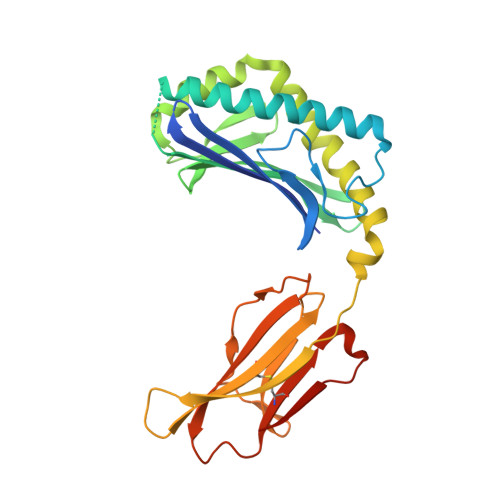
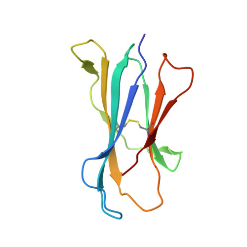
Design of natural killer T cell activators: structure and function of a microbial glycosphingolipid bound to mouse CD1d.

Wu, D.Zajonc, D.M.Fujio, M.Sullivan, B.A.Kinjo, Y.Kronenberg, M.Wilson, I.A.Wong, C.H.

(2006) Proc Natl Acad Sci U S A 103: 3972-3977

  • DOI: https://doi.org/10.1073/pnas.0600285103
  • Primary Citation of Related Structures:  
    2FIK

  • PubMed Abstract: 

    Natural killer T (NKT) cells provide an innate-type immune response upon T cell receptor interaction with CD1d-presented antigens. We demonstrate through equilibrium tetramer binding and antigen presentation assays with Valpha14i-positive NKT cell hybridomas that the Sphingomonas glycolipid alpha-galacturonosyl ceramide (GalA-GSL) is a NKT cell agonist that is significantly weaker than alpha-galactosylceramide (alpha-GalCer), the most potent known NKT agonist. For GalA-GSL, a shorter fatty acyl chain, an absence of the 4-OH on the sphingosine tail and a 6'-COOH group on the galactose moiety account for its observed antigenic potency. We further determined the crystal structure of mCD1d in complex with GalA-GSL at 1.8-A resolution. The overall binding mode of GalA-GSL to mCD1d is similar to that of the short-chain alpha-GalCer ligand PBS-25, but its sphinganine chain is more deeply inserted into the F' pocket due to alternate hydrogen-bonding interactions between the sphinganine 3-OH with Asp-80. Subsequently, a slight lateral shift (>1 A) of the galacturonosyl head group is noted at the CD1 surface compared with the galactose of alpha-GalCer. Because the relatively short C(14) fatty acid of GalA-GSL does not fully occupy the A' pocket, a spacer lipid is found that stabilizes this pocket. The lipid spacer was identified by GC/MS as a mixture of saturated and monounsaturated palmitic acid (C(16)). Comparison of available crystal structures of alpha-anomeric glycosphingolipids now sheds light on the structural basis of their differential antigenic potency and has led to the design and synthesis of NKT cell agonists with enhanced cell-based stimulatory activities compared with alpha-GalCer.


  • Organizational Affiliation

    Department of Chemistry and Molecular Biology and The Skaggs Institute for Chemical Biology, The Scripps Research Institute, 10550 North Torrey Pines Road, La Jolla, CA 92037, USA.


Macromolecules
Find similar proteins by:  (by identity cutoff)  |  3D Structure
Entity ID: 1
MoleculeChains Sequence LengthOrganismDetailsImage
T-cell surface glycoprotein CD1d1287Mus musculusMutation(s): 0 
Gene Names: Cd1d1Cd1.1
UniProt & NIH Common Fund Data Resources
Find proteins for P11609 (Mus musculus)
Explore P11609 
Go to UniProtKB:  P11609
IMPC:  MGI:107674
Entity Groups  
Sequence Clusters30% Identity50% Identity70% Identity90% Identity95% Identity100% Identity
UniProt GroupP11609
Sequence Annotations
Expand
  • Reference Sequence
Find similar proteins by:  (by identity cutoff)  |  3D Structure
Entity ID: 2
MoleculeChains Sequence LengthOrganismDetailsImage
B(2)-microglobulin99Mus musculusMutation(s): 0 
Gene Names: beta-2-microglobulin
UniProt & NIH Common Fund Data Resources
Find proteins for P01887 (Mus musculus)
Explore P01887 
Go to UniProtKB:  P01887
IMPC:  MGI:88127
Entity Groups  
Sequence Clusters30% Identity50% Identity70% Identity90% Identity95% Identity100% Identity
UniProt GroupP01887
Sequence Annotations
Expand
  • Reference Sequence
Oligosaccharides

Help

Entity ID: 3
MoleculeChains Length2D Diagram Glycosylation3D Interactions
2-acetamido-2-deoxy-beta-D-glucopyranose-(1-4)-2-acetamido-2-deoxy-beta-D-glucopyranose
C
2N-Glycosylation
Glycosylation Resources
GlyTouCan:  G42666HT
GlyCosmos:  G42666HT
GlyGen:  G42666HT
Small Molecules
Ligands 3 Unique
IDChains Name / Formula / InChI Key2D Diagram3D Interactions
GSL
Query on GSL

Download Ideal Coordinates CCD File 
F [auth A](2S,3R)-3-HYDROXY-2-(TETRADECANOYLAMINO)OCTADECYL ALPHA-D-GALACTOPYRANOSIDURONIC ACID
C38 H73 N O9
IRPOZWRRAFKYMQ-LMIAXWKISA-N
PLM
Query on PLM

Download Ideal Coordinates CCD File 
G [auth A]PALMITIC ACID
C16 H32 O2
IPCSVZSSVZVIGE-UHFFFAOYSA-N
NAG
Query on NAG

Download Ideal Coordinates CCD File 
D [auth A],
E [auth A]
2-acetamido-2-deoxy-beta-D-glucopyranose
C8 H15 N O6
OVRNDRQMDRJTHS-FMDGEEDCSA-N
Experimental Data & Validation

Experimental Data

  • Method: X-RAY DIFFRACTION
  • Resolution: 1.80 Å
  • R-Value Free: 0.243 
  • R-Value Work: 0.208 
  • R-Value Observed: 0.209 
  • Space Group: P 21 21 21
Unit Cell:
Length ( Å )Angle ( ˚ )
a = 42.295α = 90
b = 107.661β = 90
c = 110.699γ = 90
Software Package:
Software NamePurpose
REFMACrefinement
MOSFLMdata reduction
CCP4data scaling
MOLREPphasing

Structure Validation

View Full Validation Report


Ligand Structure Quality Assessment 


Entry History 

Deposition Data

Revision History  (Full details and data files)

  • Version 1.0: 2006-03-21
    Type: Initial release
  • Version 1.1: 2008-05-01
    Changes: Version format compliance
  • Version 1.2: 2011-07-13
    Changes: Non-polymer description, Version format compliance
  • Version 2.0: 2020-07-29
    Type: Remediation
    Reason: Carbohydrate remediation
    Changes: Advisory, Atomic model, Data collection, Database references, Derived calculations, Structure summary
  • Version 2.1: 2023-08-30
    Changes: Advisory, Data collection, Database references, Refinement description, Structure summary