2FF4

Mycobacterium tuberculosis EmbR in complex with low affinity phosphopeptide


Experimental Data Snapshot

  • Method: X-RAY DIFFRACTION
  • Resolution: 1.90 Å
  • R-Value Free: 0.239 
  • R-Value Work: 0.215 
  • R-Value Observed: 0.216 

wwPDB Validation   3D Report Full Report


This is version 1.2 of the entry. See complete history


Literature

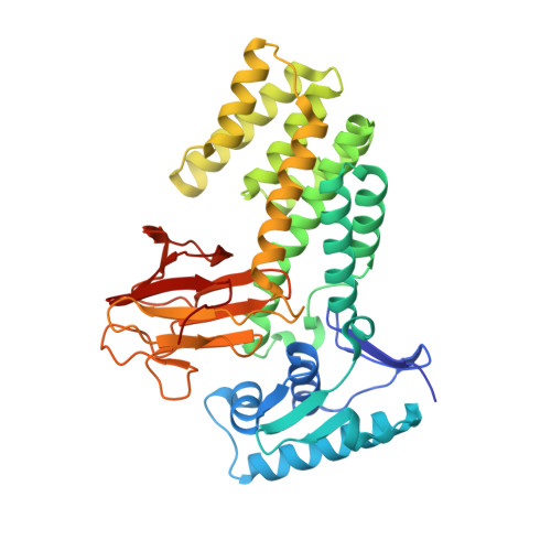
Molecular structure of EmbR, a response element of Ser/Thr kinase signaling in Mycobacterium tuberculosis.

Alderwick, L.J.Molle, V.Kremer, L.Cozzone, A.J.Dafforn, T.R.Besra, G.S.Futterer, K.

(2006) Proc Natl Acad Sci U S A 103: 2558-2563

  • DOI: https://doi.org/10.1073/pnas.0507766103
  • Primary Citation of Related Structures:  
    2FEZ, 2FF4

  • PubMed Abstract: 

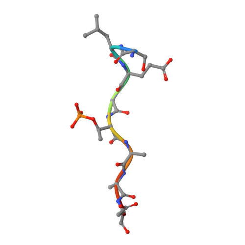
    Ser/Thr phosphorylation has emerged as a critical regulatory mechanism in a number of bacteria, including Mycobacterium tuberculosis. This problematic pathogen encodes 11 eukaryotic-like Ser/Thr kinases, yet few substrates or signaling targets have been characterized. Here, we report the structure of EmbR (2.0 A), a putative transcriptional regulator of key arabinosyltransferases (EmbC, -A, and -B), and an endogenous substrate of the Ser/Thr-kinase PknH. EmbR presents a unique domain architecture: the N-terminal winged-helix DNA-binding domain forms an extensive interface with the all-helical central bacterial transcriptional activation domain and is positioned adjacent to the regulatory C-terminal forkhead-associated (FHA) domain, which mediates binding to a Thr-phosphorylated site in PknH. The structure in complex with a phospho-peptide (1.9 A) reveals a conserved mode of phospho-threonine recognition by the FHA domain and evidence for specific recognition of the cognate kinase. The present structures suggest hypotheses as to how EmbR might propagate the phospho-relay signal from its cognate kinase, while serving as a template for the structurally uncharacterized Streptomyces antibiotic regulatory protein family of transcription factors.


  • Organizational Affiliation

    School of Biosciences, University of Birmingham, Edgbaston, Birmingham B15 2TT, United Kingdom.


Macromolecules
Find similar proteins by:  (by identity cutoff)  |  3D Structure
Entity ID: 1
MoleculeChains Sequence LengthOrganismDetailsImage
Probable regulatory protein embR
A, B
388Mycobacterium tuberculosis H37RvMutation(s): 0 
Gene Names: embR
UniProt
Find proteins for P9WGJ9 (Mycobacterium tuberculosis (strain ATCC 25618 / H37Rv))
Explore P9WGJ9 
Go to UniProtKB:  P9WGJ9
Entity Groups  
Sequence Clusters30% Identity50% Identity70% Identity90% Identity95% Identity100% Identity
UniProt GroupP9WGJ9
Sequence Annotations
Expand
  • Reference Sequence

Find similar proteins by:  Sequence   |   3D Structure  

Entity ID: 2
MoleculeChains Sequence LengthOrganismDetailsImage
DNA repair protein RAD9C [auth E],
D [auth F]
9N/AMutation(s): 1 
UniProt
Find proteins for P14737 (Saccharomyces cerevisiae (strain ATCC 204508 / S288c))
Explore P14737 
Go to UniProtKB:  P14737
Entity Groups  
UniProt GroupP14737
Sequence Annotations
Expand
  • Reference Sequence
Small Molecules
Modified Residues  1 Unique
IDChains TypeFormula2D DiagramParent
TPO
Query on TPO
C [auth E],
D [auth F]
L-PEPTIDE LINKINGC4 H10 N O6 PTHR
Experimental Data & Validation

Experimental Data

  • Method: X-RAY DIFFRACTION
  • Resolution: 1.90 Å
  • R-Value Free: 0.239 
  • R-Value Work: 0.215 
  • R-Value Observed: 0.216 
  • Space Group: C 1 2 1
Unit Cell:
Length ( Å )Angle ( ˚ )
a = 162.39α = 90
b = 82.04β = 115.37
c = 81.08γ = 90
Software Package:
Software NamePurpose
REFMACrefinement
XDSdata reduction
XDSdata scaling
PHASERphasing

Structure Validation

View Full Validation Report


Entry History 

Deposition Data

Revision History  (Full details and data files)

  • Version 1.0: 2006-01-24
    Type: Initial release
  • Version 1.1: 2008-05-01
    Changes: Version format compliance
  • Version 1.2: 2011-07-13
    Changes: Advisory, Source and taxonomy, Version format compliance