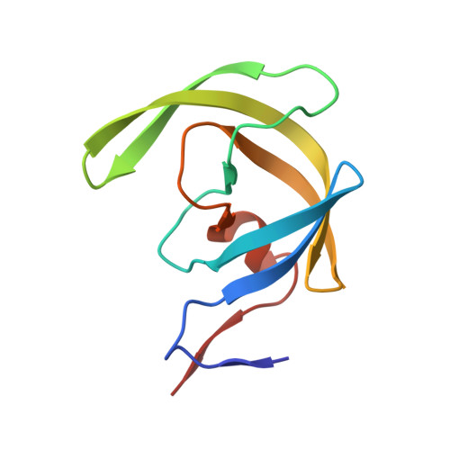
2F3K

Substrate envelope and drug resistance: crystal structure of r01 in complex with wild-type hiv-1 protease


Experimental Data Snapshot

  • Method: X-RAY DIFFRACTION
  • Resolution: 1.60 Å
  • R-Value Free: 0.197 
  • R-Value Work: 0.163 
  • R-Value Observed: 0.165 

wwPDB Validation   3D Report Full Report


Ligand Structure Quality Assessment 


This is version 1.5 of the entry. See complete history


Literature

Substrate envelope and drug resistance: crystal structure of RO1 in complex with wild-type human immunodeficiency virus type 1 protease.

Prabu-Jeyabalan, M.King, N.M.Nalivaika, E.A.Heilek-Snyder, G.Cammack, N.Schiffer, C.A.

(2006) Antimicrob Agents Chemother 50: 1518-1521

  • DOI: https://doi.org/10.1128/AAC.50.4.1518-1521.2006
  • Primary Citation of Related Structures:  
    2F3K

  • PubMed Abstract: 

    In our previous crystallographic studies of human immunodeficiency virus type 1 (HIV-1) protease-substrate complexes, we described a conserved "envelope" that appears to be important for substrate recognition and the selection of drug-resistant mutations. In this study, the complex of HIV-1 protease with the inhibitor RO1 was determined and comparison with the substrate envelope provides a rationale for mutational patterns.


  • Organizational Affiliation

    Department of Biochemistry & Molecular Pharmacology, University of Massachusetts Medical School, 364 Plantation St., Worcester, MA 01605, USA.


Macromolecules
Find similar proteins by:  (by identity cutoff)  |  3D Structure
Entity ID: 1
MoleculeChains Sequence LengthOrganismDetailsImage
Protease
A, B
99Human immunodeficiency virus 1Mutation(s): 1 
EC: 3.4.23.16
UniProt
Find proteins for O38708 (Human immunodeficiency virus 1)
Explore O38708 
Go to UniProtKB:  O38708
Entity Groups  
Sequence Clusters30% Identity50% Identity70% Identity90% Identity95% Identity100% Identity
UniProt GroupO38708
Sequence Annotations
Expand
  • Reference Sequence
Small Molecules
Binding Affinity Annotations 
IDSourceBinding Affinity
RO1 Binding MOAD:  2F3K Kd: 0.95 (nM) from 1 assay(s)
PDBBind:  2F3K IC50: 13 (nM) from 1 assay(s)
Experimental Data & Validation

Experimental Data

  • Method: X-RAY DIFFRACTION
  • Resolution: 1.60 Å
  • R-Value Free: 0.197 
  • R-Value Work: 0.163 
  • R-Value Observed: 0.165 
  • Space Group: P 21 21 21
Unit Cell:
Length ( Å )Angle ( ˚ )
a = 50.873α = 90
b = 58.167β = 90
c = 61.653γ = 90
Software Package:
Software NamePurpose
REFMACrefinement
SCALEPACKdata scaling
AMoREphasing

Structure Validation

View Full Validation Report


Ligand Structure Quality Assessment 


Entry History 

Deposition Data

Revision History  (Full details and data files)

  • Version 1.0: 2006-11-21
    Type: Initial release
  • Version 1.1: 2008-05-01
    Changes: Version format compliance
  • Version 1.2: 2011-07-13
    Changes: Version format compliance
  • Version 1.3: 2017-10-18
    Changes: Refinement description
  • Version 1.4: 2021-10-20
    Changes: Database references, Derived calculations
  • Version 1.5: 2023-08-23
    Changes: Data collection, Refinement description