


The Crystal Structure of Human Mitochondrial Acetoacetyl-Coa Thiolase Acat1.
Dombrovski, L., Min, J.R., Antoshenko, T., Wu, H., Loppnau, P., Edwards, A.M., Arrowsmith, C.H., Bochkarev, A., Plotnikov, A.N.To be published.
Experimental Data Snapshot
Entity ID: 1 | |||||
|---|---|---|---|---|---|
| Molecule | Chains | Sequence Length | Organism | Details | Image |
| Acetyl-CoA acetyltransferase, mitochondrial | 406 | Homo sapiens | Mutation(s): 1 Gene Names: ACAT1, ACAT, MAT EC: 2.3.1.9 | 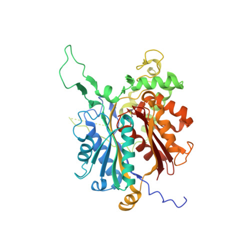 | |
UniProt & NIH Common Fund Data Resources | |||||
Find proteins for P24752 (Homo sapiens) Explore P24752 Go to UniProtKB: P24752 | |||||
PHAROS: P24752 GTEx: ENSG00000075239 | |||||
Entity Groups | |||||
| Sequence Clusters | 30% Identity50% Identity70% Identity90% Identity95% Identity100% Identity | ||||
| UniProt Group | P24752 | ||||
Sequence AnnotationsExpand | |||||
| |||||
| Ligands 2 Unique | |||||
|---|---|---|---|---|---|
| ID | Chains | Name / Formula / InChI Key | 2D Diagram | 3D Interactions | |
| COA Query on COA | F [auth A], I [auth B], J [auth C], K [auth D] | COENZYME A C21 H36 N7 O16 P3 S RGJOEKWQDUBAIZ-IBOSZNHHSA-N |  | ||
| CL Query on CL | E [auth A], G [auth B], H [auth B] | CHLORIDE ION Cl VEXZGXHMUGYJMC-UHFFFAOYSA-M |  | ||
| Modified Residues 1 Unique | |||||
|---|---|---|---|---|---|
| ID | Chains | Type | Formula | 2D Diagram | Parent |
| SCY Query on SCY | A, B, C, D | L-PEPTIDE LINKING | C5 H9 N O3 S |  | CYS |
| Length ( Å ) | Angle ( ˚ ) |
|---|---|
| a = 56.989 | α = 90 |
| b = 126.64 | β = 98.64 |
| c = 111.858 | γ = 90 |
| Software Name | Purpose |
|---|---|
| HKL-2000 | data collection |
| SCALEPACK | data scaling |
| PHASER | phasing |
| REFMAC | refinement |
| HKL-2000 | data reduction |
RCSB PDB (citation) is hosted by
RCSB PDB is a member of the

