2F19

THREE-DIMENSIONAL STRUCTURE OF TWO CRYSTAL FORMS OF FAB R19.9, FROM A MONOCLONAL ANTI-ARSONATE ANTIBODY


Experimental Data Snapshot

  • Method: X-RAY DIFFRACTION
  • Resolution: 2.80 Å
  • R-Value Work: 0.182 

wwPDB Validation   3D Report Full Report


This is version 1.2 of the entry. See complete history


Literature

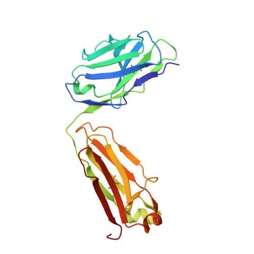
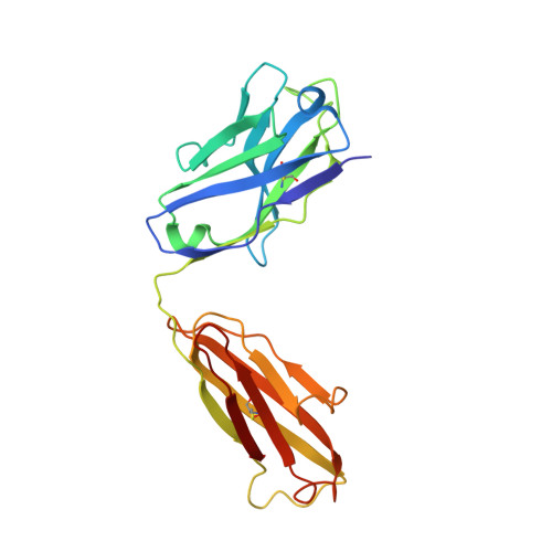
Three-dimensional structure of two crystal forms of FabR19.9 from a monoclonal anti-arsonate antibody.

Lascombe, M.-B.Alzari, P.M.Poljak, R.J.Nisonoff, A.

(1992) Proc Natl Acad Sci U S A 89: 9429-9433

  • DOI: https://doi.org/10.1073/pnas.89.20.9429
  • Primary Citation of Related Structures:  
    1FAI, 2F19

  • PubMed Abstract: 

    The three-dimensional structure of FabR19.9 from a well-characterized anti-p-azobenzenearsonate monoclonal antibody has been determined by x-ray diffraction techniques in two crystalline forms (I and II) to a resolution of 2.8 and 2.7 A, respectively. Essentially the same tertiary and quaternary structure of the Fab is observed in the two forms. The major difference resides in the intermolecular contacts, which are interpreted to favor an irreversible transition from the metastable form I to the more stable form II. The third complementarity-determining region of the heavy chain (H3) folds back over the combining site and requires rearrangement for hapten binding. This dynamic requirement on H3 is consistent with its mobility in the structure and can explain hapten binding to an otherwise inaccessible antibody combining site.


  • Organizational Affiliation

    Unite de Recherche Associée 359 Centre National de la Recherche Scientifique, Institut Pasteur, Paris, France.


Macromolecules
Find similar proteins by:  (by identity cutoff)  |  3D Structure
Entity ID: 1
MoleculeChains Sequence LengthOrganismDetailsImage
IGG2B-KAPPA R19.9 FAB (LIGHT CHAIN)A [auth L]214Mus musculusMutation(s): 0 
UniProt
Find proteins for P01837 (Mus musculus)
Explore P01837 
Go to UniProtKB:  P01837
Entity Groups  
Sequence Clusters30% Identity50% Identity70% Identity90% Identity95% Identity100% Identity
UniProt GroupP01837
Sequence Annotations
Expand
  • Reference Sequence
Find similar proteins by:  (by identity cutoff)  |  3D Structure
Entity ID: 2
MoleculeChains Sequence LengthOrganismDetailsImage
IGG2B-KAPPA R19.9 FAB (HEAVY CHAIN)B [auth H]221Mus musculusMutation(s): 0 
Entity Groups  
Sequence Clusters30% Identity50% Identity70% Identity90% Identity95% Identity100% Identity
Sequence Annotations
Expand
  • Reference Sequence
Experimental Data & Validation

Experimental Data

  • Method: X-RAY DIFFRACTION
  • Resolution: 2.80 Å
  • R-Value Work: 0.182 
  • Space Group: P 1 21 1
Unit Cell:
Length ( Å )Angle ( ˚ )
a = 43.3α = 90
b = 80.8β = 96
c = 75.1γ = 90
Software Package:
Software NamePurpose
X-PLORrefinement

Structure Validation

View Full Validation Report


Entry History 

Deposition Data

Revision History  (Full details and data files)

  • Version 1.0: 1992-10-15
    Type: Initial release
  • Version 1.1: 2008-03-24
    Changes: Version format compliance
  • Version 1.2: 2011-07-13
    Changes: Version format compliance