2ESW

Atomic structure of the N-terminal SH3 domain of mouse beta PIX,p21-activated kinase (PAK)-interacting exchange factor


Experimental Data Snapshot

  • Method: X-RAY DIFFRACTION
  • Resolution: 2.01 Å
  • R-Value Free: 0.254 
  • R-Value Work: 0.232 
  • R-Value Observed: 0.254 

wwPDB Validation   3D Report Full Report


This is version 1.3 of the entry. See complete history


Literature

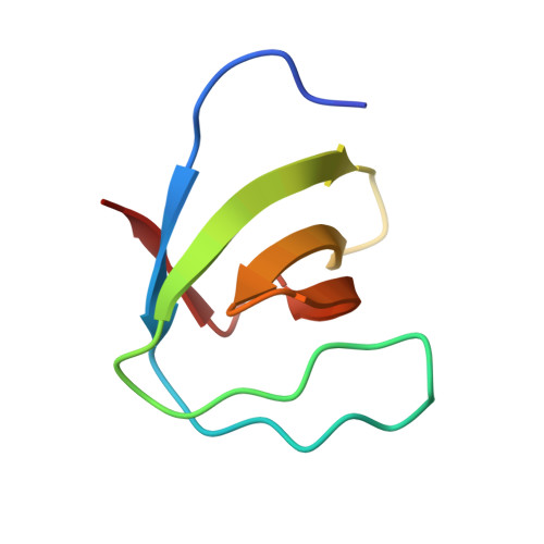
Crystal structure of the N-terminal SH3 domain of mouse betaPIX, p21-activated kinase-interacting exchange factor

Li, X.Liu, X.Sun, F.Gao, J.Zhou, H.Gao, G.F.Bartlam, M.Rao, Z.

(2006) Biochem Biophys Res Commun 339: 407-414

  • DOI: https://doi.org/10.1016/j.bbrc.2005.10.212
  • Primary Citation of Related Structures:  
    2ESW

  • PubMed Abstract: 

    The mouse betaPIX-SH3 domain, residues 8-63 of P21-activated kinase interacting exchange factor, has been characterized by X-ray diffraction. Crystals belonging to space group P3(2)21 diffracted to 2.0 A and the structure was phased by the single-wavelength anomalous diffraction method. The domain is a compact beta-barrel with an overall conformation similar to the general SH3 structure. The X-ray structure shows mouse betaPIX-SH3 domain binding the way in which the betaPIX characteristic amino acids do so for an unconventional ligand binding surface. This arrangement provides a rationale for the unusual ligand recognition motif exhibited by mouse betaPIX-SH3 domain. Comparison with another SH3/peptide complex shows that the recognition mode of the mouse betaPIX-SH3 domain should be very similar to the RXXK ligand binding mode. The unique large and planar hydrophobic pocket may contribute to the promiscuity of betaPIX-SH3 domain resulting in its multiple biological functions.


  • Organizational Affiliation

    Laboratory of Structural Biology, Tsinghua University, Beijing 100084, China.


Macromolecules
Find similar proteins by:  (by identity cutoff)  |  3D Structure
Entity ID: 1
MoleculeChains Sequence LengthOrganismDetailsImage
Rho guanine nucleotide exchange factor 7
A, B
61Mus musculusMutation(s): 0 
UniProt & NIH Common Fund Data Resources
Find proteins for Q9ES28 (Mus musculus)
Explore Q9ES28 
Go to UniProtKB:  Q9ES28
IMPC:  MGI:1860493
Entity Groups  
Sequence Clusters30% Identity50% Identity70% Identity90% Identity95% Identity100% Identity
UniProt GroupQ9ES28
Sequence Annotations
Expand
  • Reference Sequence
Experimental Data & Validation

Experimental Data

  • Method: X-RAY DIFFRACTION
  • Resolution: 2.01 Å
  • R-Value Free: 0.254 
  • R-Value Work: 0.232 
  • R-Value Observed: 0.254 
  • Space Group: P 32 2 1
Unit Cell:
Length ( Å )Angle ( ˚ )
a = 88.287α = 90
b = 88.287β = 90
c = 40.558γ = 120
Software Package:
Software NamePurpose
HKL-2000data collection
SCALEPACKdata scaling
SOLVEphasing
CNSrefinement
HKL-2000data reduction

Structure Validation

View Full Validation Report


Entry History 

Deposition Data

  • Released Date: 2006-04-11 
  • Deposition Author(s): Rao, Z.

Revision History  (Full details and data files)

  • Version 1.0: 2006-04-11
    Type: Initial release
  • Version 1.1: 2008-05-01
    Changes: Version format compliance
  • Version 1.2: 2011-07-13
    Changes: Version format compliance
  • Version 1.3: 2024-03-13
    Changes: Data collection, Database references, Derived calculations