2EBO

CORE STRUCTURE OF GP2 FROM EBOLA VIRUS


Experimental Data Snapshot

  • Method: X-RAY DIFFRACTION
  • Resolution: 1.90 Å
  • R-Value Free: 0.270 
  • R-Value Work: 0.205 
  • R-Value Observed: 0.205 

wwPDB Validation   3D Report Full Report


This is version 1.4 of the entry. See complete history


Literature

Core structure of the envelope glycoprotein GP2 from Ebola virus at 1.9-A resolution.

Malashkevich, V.N.Schneider, B.J.McNally, M.L.Milhollen, M.A.Pang, J.X.Kim, P.S.

(1999) Proc Natl Acad Sci U S A 96: 2662-2667

  • DOI: https://doi.org/10.1073/pnas.96.6.2662
  • Primary Citation of Related Structures:  
    2EBO

  • PubMed Abstract: 

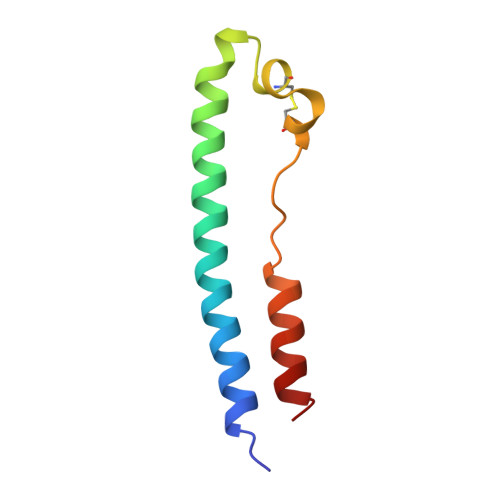
    Ebola virions contain a surface transmembrane glycoprotein (GP) that is responsible for binding to target cells and subsequent fusion of the viral and host-cell membranes. GP is expressed as a single-chain precursor that is posttranslationally processed into the disulfide-linked fragments GP1 and GP2. The GP2 subunit is thought to mediate membrane fusion. A soluble fragment of the GP2 ectodomain, lacking the fusion-peptide region and the transmembrane helix, folds into a stable, highly helical structure in aqueous solution. Limited proteolysis studies identify a stable core of the GP2 ectodomain. This 74-residue core, denoted Ebo-74, was crystallized, and its x-ray structure was determined at 1.9-A resolution. Ebo-74 forms a trimer in which a long, central three-stranded coiled coil is surrounded by shorter C-terminal helices that are packed in an antiparallel orientation into hydrophobic grooves on the surface of the coiled coil. Our results confirm the previously anticipated structural similarity between the Ebola GP2 ectodomain and the core of the transmembrane subunit from oncogenic retroviruses. The Ebo-74 structure likely represents the fusion-active conformation of the protein, and its overall architecture resembles several other viral membrane-fusion proteins, including those from HIV and influenza.


  • Organizational Affiliation

    Howard Hughes Medical Institute, Whitehead Institute for Biomedical Research, Department of Biology, Massachusetts Institute of Technology, Nine Cambridge Center, Cambridge, MA 02142, USA.


Macromolecules
Find similar proteins by:  (by identity cutoff)  |  3D Structure
Entity ID: 1
MoleculeChains Sequence LengthOrganismDetailsImage
EBOLA VIRUS ENVELOPE GLYCOPROTEIN
A, B, C
74Ebola virus - Mayinga, Zaire, 1976Mutation(s): 1 
Gene Names: GP41
UniProt
Find proteins for Q05320 (Zaire ebolavirus (strain Mayinga-76))
Explore Q05320 
Go to UniProtKB:  Q05320
Entity Groups  
Sequence Clusters30% Identity50% Identity70% Identity90% Identity95% Identity100% Identity
UniProt GroupQ05320
Sequence Annotations
Expand
  • Reference Sequence
Small Molecules
Ligands 1 Unique
IDChains Name / Formula / InChI Key2D Diagram3D Interactions
CL
Query on CL

Download Ideal Coordinates CCD File 
D [auth A]CHLORIDE ION
Cl
VEXZGXHMUGYJMC-UHFFFAOYSA-M
Experimental Data & Validation

Experimental Data

  • Method: X-RAY DIFFRACTION
  • Resolution: 1.90 Å
  • R-Value Free: 0.270 
  • R-Value Work: 0.205 
  • R-Value Observed: 0.205 
  • Space Group: P 62
Unit Cell:
Length ( Å )Angle ( ˚ )
a = 75.66α = 90
b = 75.66β = 90
c = 67.93γ = 120
Software Package:
Software NamePurpose
DENZOdata reduction
SCALEPACKdata scaling
CNSrefinement
CNSphasing

Structure Validation

View Full Validation Report


Entry History 

Deposition Data

Revision History  (Full details and data files)

  • Version 1.0: 1999-05-18
    Type: Initial release
  • Version 1.1: 2008-03-24
    Changes: Version format compliance
  • Version 1.2: 2011-07-13
    Changes: Version format compliance
  • Version 1.3: 2018-03-14
    Changes: Database references
  • Version 1.4: 2023-08-09
    Changes: Database references, Derived calculations, Refinement description