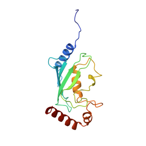
Crystal structure of the cyclin-specific ubiquitin-conjugating enzyme from clam, E2-C, at 2.0 A resolution.
Jiang, F., Basavappa, R.(1999) Biochemistry 38: 6471-6478
- PubMed: 10350465
- DOI: https://doi.org/10.1021/bi9901329
- Primary Citation of Related Structures:
2E2C - PubMed Abstract:
The destruction of the cyclin B protein is necessary for the cell to exit from mitosis. The destruction of cyclin B occurs via the ubiquitin/proteasome system and involves a specific ubiquitin-conjugating enzyme (Ubc) that donates ubiquitin to cyclin B. Here we present the crystal structure of the cyclin-specific Ubc from clam, E2-C, determined at 2.0 A resolution. The E2-C enzyme contains an N-terminal extension in addition to the Ubc core domain. The N-terminal extension is disordered, perhaps reflecting a need for flexibility as it interacts with various partners in the ubiquitination system. The overall structure of the E2-C core domain is quite similar to those in previously determined Ubc proteins. The interaction between particular pairs of E2-C proteins in the crystal has some of the hallmarks of a functional dimer, though solution studies suggest that the E2-C protein exists as a monomer. Comparison of the E2-C structure with that of the other available Ubc structures indicates conserved surface residues that may interact with common components of the ubiquitination system. Such comparison also reveals a remarkable spine of conserved hydrophobic residues in the center of the protein that may drive the protein to fold and stabilize the protein once folded. Comparison of residues conserved only among E2-C and its homologues indicates surface areas that may be involved in mitotic-specific ubiquitination.
Organizational Affiliation:
Department of Biochemistry and Biophysics, University of Rochester Medical Center, New York 14642, USA.