2DWN

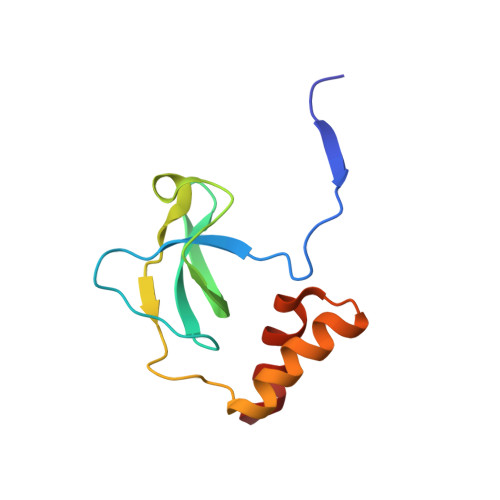
Crystal structure of the PriA protein complexed with oligonucleotides


Experimental Data Snapshot

  • Method: X-RAY DIFFRACTION
  • Resolution: 3.35 Å
  • R-Value Free: 0.333 
  • R-Value Work: 0.281 

wwPDB Validation   3D Report Full Report


This is version 2.1 of the entry. See complete history


Literature

Structural basis of the 3'-end recognition of a leading strand in stalled replication forks by PriA.

Sasaki, K.Ose, T.Okamoto, N.Maenaka, K.Tanaka, T.Masai, H.Saito, M.Shirai, T.Kohda, D.

(2007) EMBO J 26: 2584-2593

  • DOI: https://doi.org/10.1038/sj.emboj.7601697
  • Primary Citation of Related Structures:  
    2D7E, 2D7G, 2D7H, 2DWL, 2DWM, 2DWN

  • PubMed Abstract: 

    In eubacteria, PriA helicase detects the stalled DNA replication forks. This critical role of PriA is ascribed to its ability to bind to the 3' end of a nascent leading DNA strand in the stalled replication forks. The crystal structures in complexes with oligonucleotides and the combination of fluorescence correlation spectroscopy and mutagenesis reveal that the N-terminal domain of PriA possesses a binding pocket for the 3'-terminal nucleotide residue of DNA. The interaction with the deoxyribose 3'-OH is essential for the 3'-terminal recognition. In contrast, the direct interaction with 3'-end nucleobase is unexpected, considering the same affinity for oligonucleotides carrying the four bases at the 3' end. Thus, the N-terminal domain of PriA recognizes the 3'-end base in a base-non-selective manner, in addition to the deoxyribose and 5'-side phosphodiester group, of the 3'-terminal nucleotide to acquire both sufficient affinity and non-selectivity to find all of the stalled replication forks generated during DNA duplication. This unique feature is prerequisite for the proper positioning of the helicase domain of PriA on the unreplicated double-stranded DNA.


  • Organizational Affiliation

    Division of Structural Biology, Medical Institute of Bioregulation, Kyushu University, Fukuoka, Japan.


Macromolecules

Find similar proteins by:  (by identity cutoff)  |  3D Structure
Entity ID: 1
MoleculeChains Sequence LengthOrganismDetailsImage
Primosomal protein N'
A, B, C, D
105Escherichia coli K-12Mutation(s): 0 
Gene Names: priAb3935JW3906
EC: 3.6.4
UniProt
Find proteins for P17888 (Escherichia coli (strain K12))
Explore P17888 
Go to UniProtKB:  P17888
Entity Groups  
Sequence Clusters30% Identity50% Identity70% Identity90% Identity95% Identity100% Identity
UniProt GroupP17888
Sequence Annotations
Expand
  • Reference Sequence

Find similar nucleic acids by:  Sequence   |   3D Structure  

Entity ID: 2
MoleculeChains LengthOrganismImage
DNA (5'-D(*A*G)-3')
E, F
2synthetic construct
Sequence Annotations
Expand
  • Reference Sequence
Experimental Data & Validation

Experimental Data

  • Method: X-RAY DIFFRACTION
  • Resolution: 3.35 Å
  • R-Value Free: 0.333 
  • R-Value Work: 0.281 
  • Space Group: H 3 2
Unit Cell:
Length ( Å )Angle ( ˚ )
a = 112.25α = 90
b = 112.25β = 90
c = 260.83γ = 120
Software Package:
Software NamePurpose
HKL-2000data collection
MOLREPphasing
CNSrefinement
HKL-2000data reduction
SCALEPACKdata scaling

Structure Validation

View Full Validation Report


Entry History 

Deposition Data

Revision History  (Full details and data files)

  • Version 1.0: 2006-11-07
    Type: Initial release
  • Version 1.1: 2008-04-30
    Changes: Version format compliance
  • Version 1.2: 2011-07-13
    Changes: Derived calculations, Version format compliance
  • Version 1.3: 2019-02-27
    Changes: Data collection, Database references, Source and taxonomy, Structure summary
  • Version 2.0: 2019-03-13
    Changes: Advisory, Atomic model, Data collection, Derived calculations, Non-polymer description, Polymer sequence, Structure summary
  • Version 2.1: 2023-10-25
    Changes: Data collection, Database references, Refinement description