2DRP

THE CRYSTAL STRUCTURE OF A TWO ZINC-FINGER PEPTIDE REVEALS AN EXTENSION TO THE RULES FOR ZINC-FINGER/DNA RECOGNITION


Experimental Data Snapshot

  • Method: X-RAY DIFFRACTION
  • Resolution: 2.80 Å
  • R-Value Work: 0.193 
  • R-Value Observed: 0.193 

wwPDB Validation   3D Report Full Report


This is version 1.3 of the entry. See complete history


Literature

The crystal structure of a two zinc-finger peptide reveals an extension to the rules for zinc-finger/DNA recognition.

Fairall, L.Schwabe, J.W.Chapman, L.Finch, J.T.Rhodes, D.

(1993) Nature 366: 483-487

  • DOI: https://doi.org/10.1038/366483a0
  • Primary Citation of Related Structures:  
    2DRP

  • PubMed Abstract: 

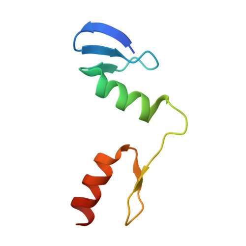
    The Cys2-His2 zinc-finger is the most widely occurring DNA-binding motif. The first structure of a zinc-finger/DNA complex revealed a fairly simple mechanism for DNA recognition suggesting that the zinc-finger might represent a candidate template for designing proteins to recognize DNA. Residues at three key positions in an alpha-helical 'reading head' play a dominant role in base-recognition and have been targets for mutagenesis experiments aimed at deriving a recognition code. Here we report the structure of a two zinc-finger DNA-binding domain from the protein Tramtrack complexed with DNA. The amino-terminal zinc-finger and its interaction with DNA illustrate several novel features. These include the use of a serine residue, which is semi-conserved and located outside the three key positions, to make a base contact. Its role in base-recognition correlates with a large, local, protein-induced deformation of the DNA helix at a flexible A-T-A sequence and may give insight into previous mutagenesis experiments. It is apparent from this structure that zinc-finger/DNA recognition is more complex than was originally perceived.


  • Organizational Affiliation

    MRC Laboratory of Molecular Biology, Cambridge, UK.


Macromolecules

Find similar proteins by:  (by identity cutoff)  |  3D Structure
Entity ID: 3
MoleculeChains Sequence LengthOrganismDetailsImage
PROTEIN (TRAMTRACK DNA-BINDING DOMAIN)E [auth A],
F [auth D]
66Drosophila melanogasterMutation(s): 0 
UniProt
Find proteins for P17789 (Drosophila melanogaster)
Explore P17789 
Go to UniProtKB:  P17789
Entity Groups  
Sequence Clusters30% Identity50% Identity70% Identity90% Identity95% Identity100% Identity
UniProt GroupP17789
Sequence Annotations
Expand
  • Reference Sequence

Find similar nucleic acids by:  Sequence   |   3D Structure  

Entity ID: 1
MoleculeChains LengthOrganismImage
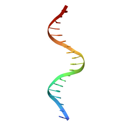
DNA (5'-D(*CP*TP*AP*AP*TP*AP*AP*GP*GP*AP*TP*AP*AP*CP*GP*TP*C P*CP*G)-3')A [auth B],
C [auth E]
19N/A
Sequence Annotations
Expand
  • Reference Sequence

Find similar nucleic acids by:  Sequence   |   3D Structure  

Entity ID: 2
MoleculeChains LengthOrganismImage
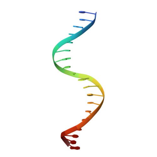
DNA (5'-D(*TP*CP*GP*GP*AP*CP*GP*TP*TP*AP*TP*CP*CP*TP*TP*AP*T P*TP*A)-3')B [auth C],
D [auth F]
19N/A
Sequence Annotations
Expand
  • Reference Sequence
Experimental Data & Validation

Experimental Data

  • Method: X-RAY DIFFRACTION
  • Resolution: 2.80 Å
  • R-Value Work: 0.193 
  • R-Value Observed: 0.193 
  • Space Group: P 21 21 21
Unit Cell:
Length ( Å )Angle ( ˚ )
a = 60.7α = 90
b = 64.6β = 90
c = 117.6γ = 90
Software Package:
Software NamePurpose
X-PLORrefinement

Structure Validation

View Full Validation Report


Entry History 

Deposition Data

Revision History  (Full details and data files)

  • Version 1.0: 1994-08-31
    Type: Initial release
  • Version 1.1: 2008-05-22
    Changes: Version format compliance
  • Version 1.2: 2011-07-13
    Changes: Version format compliance
  • Version 1.3: 2024-02-14
    Changes: Data collection, Database references, Derived calculations