2DRI

PROBING PROTEIN-PROTEIN INTERACTIONS: THE RIBOSE BINDING PROTEIN IN BACTERIAL TRANSPORT AND CHEMOTAXIS


Experimental Data Snapshot

  • Method: X-RAY DIFFRACTION
  • Resolution: 1.60 Å
  • R-Value Work: 0.194 
  • R-Value Observed: 0.194 

wwPDB Validation   3D Report Full Report


Ligand Structure Quality Assessment 


This is version 1.5 of the entry. See complete history


Literature

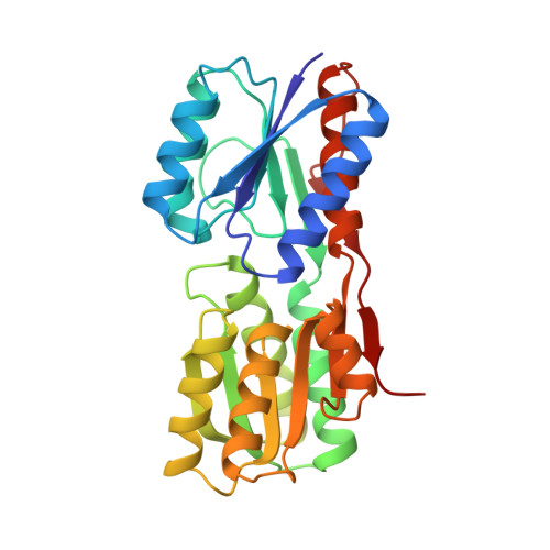
Probing protein-protein interactions. The ribose-binding protein in bacterial transport and chemotaxis.

Bjorkman, A.J.Binnie, R.A.Zhang, H.Cole, L.B.Hermodson, M.A.Mowbray, S.L.

(1994) J Biol Chem 269: 30206-30211

  • Primary Citation of Related Structures:  
    1DRJ, 1DRK, 2DRI

  • PubMed Abstract: 

    A number of mutations at Gly134 of the periplasmic ribose-binding protein of Escherichia coli were examined by a combined biochemical and structural approach. Different mutations gave rise to different patterns of effects on the chemotaxis and transport functions. The smallest residue (alanine) had the least effect on transport, whereas large hydrophobic residues had the smallest effect on chemotaxis. Comparison of the x-ray crystal structure of the G134R mutant protein (2.5-A resolution) to that of the wild type (1.6-A resolution) showed that the basic structure of the protein was unaltered. The loss of chemotaxis and transport functions in this and similar mutant proteins must therefore be caused by relatively simple surface effects, which include a change in local main chain conformation. The loss of chemotaxis and transport functions resulting from the introduction of an alanine residue at position 134 was suppressed by an additional isoleucine to threonine mutation at residue 132. An x-ray structure of the I132T/G134A double mutant protein (2.2-A resolution) showed that the changes in local structure were accompanied by a diffuse pattern of structural changes in the surrounding region, implying that the suppression derives from a combination of sources.


  • Organizational Affiliation

    Department of Molecular Biology, Swedish University of Agricultural Sciences, Uppsala.


Macromolecules
Find similar proteins by:  (by identity cutoff)  |  3D Structure
Entity ID: 1
MoleculeChains Sequence LengthOrganismDetailsImage
D-RIBOSE-BINDING PROTEIN271Escherichia coliMutation(s): 0 
UniProt
Find proteins for P02925 (Escherichia coli (strain K12))
Explore P02925 
Go to UniProtKB:  P02925
Entity Groups  
Sequence Clusters30% Identity50% Identity70% Identity90% Identity95% Identity100% Identity
UniProt GroupP02925
Sequence Annotations
Expand
  • Reference Sequence
Small Molecules
Ligands 1 Unique
IDChains Name / Formula / InChI Key2D Diagram3D Interactions
RIP
Query on RIP

Download Ideal Coordinates CCD File 
B [auth A]beta-D-ribopyranose
C5 H10 O5
SRBFZHDQGSBBOR-TXICZTDVSA-N
Binding Affinity Annotations 
IDSourceBinding Affinity
RIP Binding MOAD:  2DRI Kd: 130 (nM) from 1 assay(s)
PDBBind:  2DRI Kd: 130 (nM) from 1 assay(s)
Experimental Data & Validation

Experimental Data

  • Method: X-RAY DIFFRACTION
  • Resolution: 1.60 Å
  • R-Value Work: 0.194 
  • R-Value Observed: 0.194 
  • Space Group: P 21 21 21
Unit Cell:
Length ( Å )Angle ( ˚ )
a = 74.92α = 90
b = 88.99β = 90
c = 40.27γ = 90
Software Package:
Software NamePurpose
X-PLORmodel building
X-PLORrefinement
X-PLORphasing

Structure Validation

View Full Validation Report


Ligand Structure Quality Assessment 


Entry History 

Deposition Data

Revision History  (Full details and data files)

  • Version 1.0: 1995-01-26
    Type: Initial release
  • Version 1.1: 2008-03-03
    Changes: Version format compliance
  • Version 1.2: 2011-07-13
    Changes: Version format compliance
  • Version 1.3: 2017-11-29
    Changes: Derived calculations, Other
  • Version 1.4: 2020-07-29
    Type: Remediation
    Reason: Carbohydrate remediation
    Changes: Data collection, Derived calculations, Structure summary
  • Version 1.5: 2024-02-14
    Changes: Data collection, Database references, Structure summary