


Crystal structure of native Trypanosoma cruzi dihydroorotate dehydrogenase
Inaoka, D.K., Shimizu, H., Sakamoto, K., Shiba, T., Kurisu, G., Nara, T., Aoki, T., Harada, S., Kita, K.To be published.
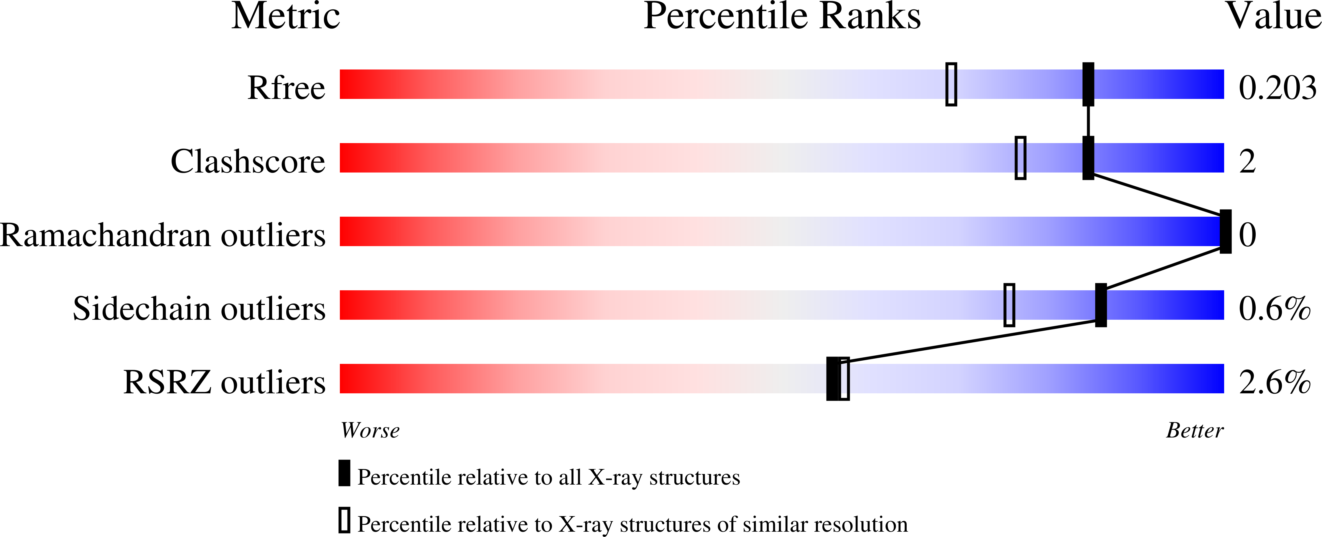
Experimental Data Snapshot
Entity ID: 1 | |||||
|---|---|---|---|---|---|
| Molecule | Chains | Sequence Length | Organism | Details | Image |
| Dihydroorotate Dehydrogenase | 314 | Trypanosoma cruzi | Mutation(s): 0 Gene Names: TCDHOD2 EC: 1.3.3.1 |  | |
UniProt | |||||
Find proteins for Q4D3W2 (Trypanosoma cruzi (strain CL Brener)) Explore Q4D3W2 Go to UniProtKB: Q4D3W2 | |||||
Entity Groups | |||||
| Sequence Clusters | 30% Identity50% Identity70% Identity90% Identity95% Identity100% Identity | ||||
| UniProt Group | Q4D3W2 | ||||
Sequence AnnotationsExpand | |||||
| |||||
| Ligands 2 Unique | |||||
|---|---|---|---|---|---|
| ID | Chains | Name / Formula / InChI Key | 2D Diagram | 3D Interactions | |
| FMN Query on FMN | D [auth A], E [auth B] | FLAVIN MONONUCLEOTIDE C17 H21 N4 O9 P FVTCRASFADXXNN-SCRDCRAPSA-N |  | ||
| NCO Query on NCO | C [auth A] | COBALT HEXAMMINE(III) Co H18 N6 DYLMFCCYOUSRTK-UHFFFAOYSA-N |  | ||
| Length ( Å ) | Angle ( ˚ ) |
|---|---|
| a = 69.959 | α = 90 |
| b = 73.128 | β = 90 |
| c = 126.087 | γ = 90 |
| Software Name | Purpose |
|---|---|
| REFMAC | refinement |
| HKL-2000 | data reduction |
| HKL-2000 | data scaling |
| MOLREP | phasing |
RCSB PDB (citation) is hosted by
RCSB PDB is a member of the

