2D7C

Crystal structure of human Rab11 in complex with FIP3 Rab-binding domain


Experimental Data Snapshot

  • Method: X-RAY DIFFRACTION
  • Resolution: 1.75 Å
  • R-Value Free: 0.224 
  • R-Value Work: 0.202 
  • R-Value Observed: 0.203 

wwPDB Validation   3D Report Full Report


Ligand Structure Quality Assessment 


This is version 1.3 of the entry. See complete history


Literature

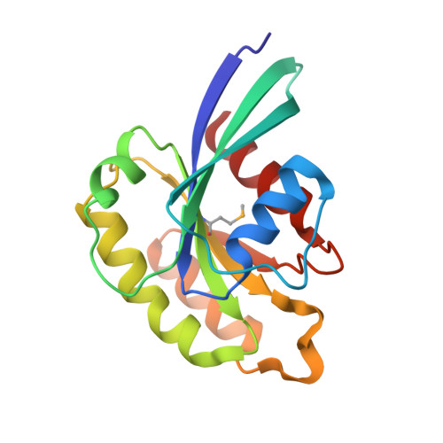
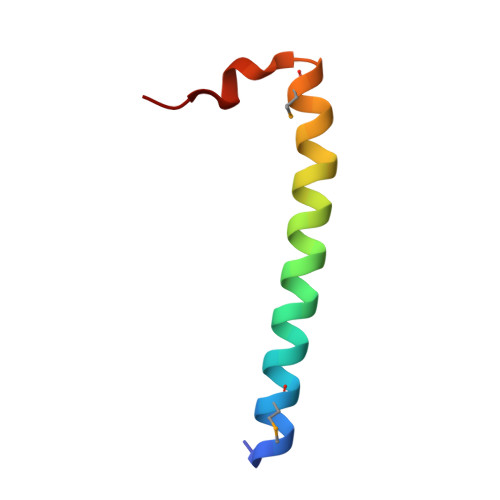
Structural basis for Rab11-dependent membrane recruitment of a family of Rab11-interacting protein 3 (FIP3)/Arfophilin-1.

Shiba, T.Koga, H.Shin, H.W.Kawasaki, M.Kato, R.Nakayama, K.Wakatsuki, S.

(2006) Proc Natl Acad Sci U S A 103: 15416-15421

  • DOI: https://doi.org/10.1073/pnas.0605357103
  • Primary Citation of Related Structures:  
    2D7C

  • PubMed Abstract: 

    Family of Rab11-interacting protein (FIP)3/Arfophlin-1 and FIP4/Arfophilin-2 are dual effectors for Rab11 and ADP ribosylation factor (ARF)5/ARF6, which are involved in membrane delivery from recycling endosomes to the plasma membrane during cytokinesis. Here, we define the distinct C-terminal binding regions of FIP3 and FIP4 for Rab11 and ARF5/ARF6. Furthermore, we determined the crystal structure of Rab11 in complex with the Rab11-binding domain (RBD) of FIP3. The long amphiphilic alpha-helix of FIP3-RBD forms a parallel coiled-coil homodimer, with two symmetric interfaces with two Rab11 molecules. The hydrophobic side of the RBD helix is involved in homodimerization and mediates the interaction with the Rab11 switch 1 region, whereas the opposite hydrophilic side interacts with the Rab11 switch 2 and is the major factor contributing to the binding specificity. The bivalent interaction of FIP3 with Rab11 at the C terminus allows FIP3 to coordinately function with other binding partners, including ARFs.


  • Organizational Affiliation

    Structural Biology Research Center, Photon Factory, Institute of Materials Structure Science, High Energy Accelerator Research Organization, Tsukuba, Ibaraki 305-0801, Japan.


Macromolecules
Find similar proteins by:  (by identity cutoff)  |  3D Structure
Entity ID: 1
MoleculeChains Sequence LengthOrganismDetailsImage
Ras-related protein Rab-11A
A, B
167Homo sapiensMutation(s): 1 
UniProt & NIH Common Fund Data Resources
Find proteins for P62491 (Homo sapiens)
Explore P62491 
Go to UniProtKB:  P62491
PHAROS:  P62491
GTEx:  ENSG00000103769 
Entity Groups  
Sequence Clusters30% Identity50% Identity70% Identity90% Identity95% Identity100% Identity
UniProt GroupP62491
Sequence Annotations
Expand
  • Reference Sequence
Find similar proteins by:  (by identity cutoff)  |  3D Structure
Entity ID: 2
MoleculeChains Sequence LengthOrganismDetailsImage
Rab11 family-interacting protein 3
C, D
42Homo sapiensMutation(s): 0 
UniProt & NIH Common Fund Data Resources
Find proteins for O75154 (Homo sapiens)
Explore O75154 
Go to UniProtKB:  O75154
PHAROS:  O75154
GTEx:  ENSG00000090565 
Entity Groups  
Sequence Clusters30% Identity50% Identity70% Identity90% Identity95% Identity100% Identity
UniProt GroupO75154
Sequence Annotations
Expand
  • Reference Sequence
Experimental Data & Validation

Experimental Data

  • Method: X-RAY DIFFRACTION
  • Resolution: 1.75 Å
  • R-Value Free: 0.224 
  • R-Value Work: 0.202 
  • R-Value Observed: 0.203 
  • Space Group: P 21 21 2
Unit Cell:
Length ( Å )Angle ( ˚ )
a = 108.717α = 90
b = 120.565β = 90
c = 36.249γ = 90
Software Package:
Software NamePurpose
HKL-2000data collection
HKL-2000data reduction
SOLVEphasing
CNSrefinement
HKL-2000data scaling

Structure Validation

View Full Validation Report


Ligand Structure Quality Assessment 


Entry History 

Deposition Data

Revision History  (Full details and data files)

  • Version 1.0: 2006-09-26
    Type: Initial release
  • Version 1.1: 2008-04-30
    Changes: Version format compliance
  • Version 1.2: 2011-07-13
    Changes: Version format compliance
  • Version 1.3: 2021-11-10
    Changes: Database references, Derived calculations