2CWG

CRYSTALLOGRAPHIC REFINEMENT AND STRUCTURE ANALYSIS OF THE COMPLEX OF WHEAT GERM AGGLUTININ WITH A BIVALENT SIALOGLYCOPEPTIDE FROM GLYCOPHORIN A


Experimental Data Snapshot

  • Method: X-RAY DIFFRACTION
  • Resolution: 2.00 Å
  • R-Value Work: 0.171 
  • R-Value Observed: 0.171 

wwPDB Validation   3D Report Full Report


This is version 4.0 of the entry. See complete history


Literature

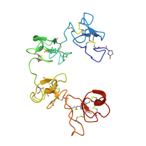
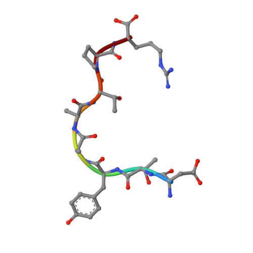
Crystallographic refinement and structure analysis of the complex of wheat germ agglutinin with a bivalent sialoglycopeptide from glycophorin A.

Wright, C.S.Jaeger, J.

(1993) J Mol Biol 232: 620-638

  • DOI: https://doi.org/10.1006/jmbi.1993.1415
  • Primary Citation of Related Structures:  
    2CWG

  • PubMed Abstract: 

    Wheat germ agglutinin (WGA) elicits a number of biological effects in erythrocytes as a result of specific binding to the transmembrane protein glycophorin A. The structure of co-crystals of WGA (isolectin 1: WGA1) with a bivalent sialoglycopeptide fragment of glycophorin A (T5), determined at 2.0 A resolution, has been further refined and analyzed with respect to ligand-induced changes in the tertiary structure, mobility, solvation, saccharide conformation and protein/saccharide interactions at three independent N-acetyl-D-neuraminic (NeuNAc) binding sites. The final model, which includes the two independent WGA1 monomers (composed of domains A, B, C and D), two positions for bound T5 sialo-tetrasaccharide (NeuNAc-alpha 2,3-Gal-beta 1,3-(alpha 2,6-NeuNAc)GalNAc) and 386 water molecules, refined to a crystallographic R-factor of 17.1% (Fo > 1.0 sigma) and an average temperature factor of 31.99 A2. Comparisons between the tertiary structures of the liganded and unliganded WGA1 dimers indicate that the largest deviations from 2-fold symmetry are localized in domains engaged in sugar binding (B1 and C2) and at the C-terminal domain of monomer II (D2), forming a strong lattice contact. Interactions of the tetrasaccharide with amino acid ligands in the three binding sites and with water were carefully analyzed and compared. Bound conformations of terminal NeuNAc match to within a root-mean-square delta r of 0.3 A. The specificity-determining N-acetyl group superimposes best in comparison with other substituents of the sugar ring. Of the five domain binding sites that are not occupied in this dimeric crosslinked complex, only one is accessible to the NeuNAc monosaccharide as determined from a difference Fourier map at 3.0 A resolution.


  • Organizational Affiliation

    Department of Biochemistry, Medical College of Virginia, Virginia Commonwealth University, Richmond 23298.


Macromolecules
Find similar proteins by:  (by identity cutoff)  |  3D Structure
Entity ID: 1
MoleculeChains Sequence LengthOrganismDetailsImage
AGGLUTININ ISOLECTIN 1 (WGA1)A,
C [auth B]
171Triticum aestivumMutation(s): 0 
UniProt
Find proteins for P10968 (Triticum aestivum)
Explore P10968 
Go to UniProtKB:  P10968
Entity Groups  
Sequence Clusters30% Identity50% Identity70% Identity90% Identity95% Identity100% Identity
UniProt GroupP10968
Sequence Annotations
Expand
  • Reference Sequence

Find similar proteins by:  Sequence   |   3D Structure  

Entity ID: 2
MoleculeChains Sequence LengthOrganismDetailsImage
T5 SIALOGLYCOPEPTIDE OF GLYCOPHORIN AB [auth D],
D [auth E]
8N/AMutation(s): 0 
Sequence Annotations
Expand
  • Reference Sequence
Oligosaccharides

Help

Entity ID: 3
MoleculeChains Length2D Diagram Glycosylation3D Interactions
N-acetyl-alpha-neuraminic acid-(2-3)-beta-D-galactopyranose-(1-3)-[N-acetyl-alpha-neuraminic acid-(2-6)]2-acetamido-2-deoxy-alpha-D-glucopyranoseE [auth C],
F
4N/A
Glycosylation Resources
GlyTouCan:  G40274ML
GlyCosmos:  G40274ML
GlyGen:  G40274ML
Small Molecules
Modified Residues  1 Unique
IDChains TypeFormula2D DiagramParent
PCA
Query on PCA
A,
C [auth B]
L-PEPTIDE LINKINGC5 H7 N O3GLN
Experimental Data & Validation

Experimental Data

  • Method: X-RAY DIFFRACTION
  • Resolution: 2.00 Å
  • R-Value Work: 0.171 
  • R-Value Observed: 0.171 
  • Space Group: P 21 21 2
Unit Cell:
Length ( Å )Angle ( ˚ )
a = 111α = 90
b = 50.4β = 90
c = 63.4γ = 90
Software Package:
Software NamePurpose
X-PLORmodel building
PROLSQrefinement
X-PLORrefinement
X-PLORphasing

Structure Validation

View Full Validation Report


Entry History 

Deposition Data

  • Released Date: 1994-01-31 
  • Deposition Author(s): Wright, C.S.

Revision History  (Full details and data files)

  • Version 1.0: 1994-01-31
    Type: Initial release
  • Version 1.1: 2008-03-03
    Changes: Version format compliance
  • Version 1.2: 2011-07-13
    Changes: Version format compliance
  • Version 1.3: 2017-11-29
    Changes: Advisory, Derived calculations, Other
  • Version 1.4: 2019-07-17
    Changes: Data collection, Derived calculations, Refinement description
  • Version 1.5: 2019-08-14
    Changes: Data collection, Refinement description
  • Version 2.0: 2019-12-25
    Changes: Data collection, Derived calculations, Polymer sequence
  • Version 3.0: 2020-04-08
    Changes: Advisory, Atomic model, Data collection, Derived calculations
  • Version 4.0: 2020-07-29
    Type: Remediation
    Reason: Carbohydrate remediation
    Changes: Advisory, Atomic model, Data collection, Derived calculations, Structure summary