2CT9

The crystal structure of calcineurin B homologous proein 1 (CHP1)


Experimental Data Snapshot

  • Method: X-RAY DIFFRACTION
  • Resolution: 2.20 Å
  • R-Value Free: 0.268 
  • R-Value Work: 0.216 
  • R-Value Observed: 0.219 

wwPDB Validation   3D Report Full Report


This is version 1.3 of the entry. See complete history


Literature

Structural characterization of calcineurin B homologous protein 1

Naoe, Y.Arita, K.Hashimoto, H.Kanazawa, H.Sato, M.Shimizu, T.

(2005) J Biol Chem 280: 32372-32378

  • DOI: https://doi.org/10.1074/jbc.M503390200
  • Primary Citation of Related Structures:  
    2CT9

  • PubMed Abstract: 

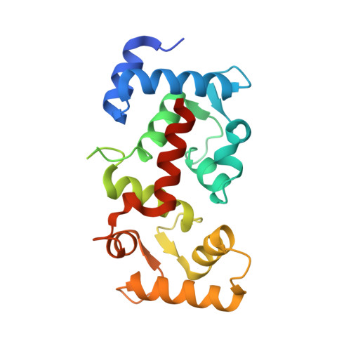
    Calcineurin B homologous protein 1 (CHP1), also known as p22, is a calcium-binding EF-hand protein that plays a role in membrane trafficking. It binds to multiple effector proteins, including Na(+)/H(+) exchangers, a serine/threonine kinase, and calcineurin, potentially modulating their function. The crystal structure of calcium-bound CHP1 from rat has been determined at 2.2 Angstroms of resolution. The molecule has a compact alpha-helical structure containing four EF-hands. The overall folding topology of the protein is similar to that of the regulatory B subunit of calcineurin and to that of calcium- and integrin-binding protein. The calcium ion is coordinated in typical fashion in the third and fourth EF-hands, but the first and second EF-hands contain no calcium ion. The first EF-hand is maintained by internal interactions, and the second EF-hand is stabilized by hydrophobic interactions. CHP1 contains a hydrophobic pocket on the opposite side of the protein to the EF-hands that has been implicated in ligand binding.


  • Organizational Affiliation

    International Graduate School of Arts and Sciences, Yokohama City University, Tsurumi-ku, Japan.


Macromolecules
Find similar proteins by:  (by identity cutoff)  |  3D Structure
Entity ID: 1
MoleculeChains Sequence LengthOrganismDetailsImage
Calcium-binding protein p22
A, B
208Rattus norvegicusMutation(s): 0 
UniProt
Find proteins for P61023 (Rattus norvegicus)
Explore P61023 
Go to UniProtKB:  P61023
Entity Groups  
Sequence Clusters30% Identity50% Identity70% Identity90% Identity95% Identity100% Identity
UniProt GroupP61023
Sequence Annotations
Expand
  • Reference Sequence
Experimental Data & Validation

Experimental Data

  • Method: X-RAY DIFFRACTION
  • Resolution: 2.20 Å
  • R-Value Free: 0.268 
  • R-Value Work: 0.216 
  • R-Value Observed: 0.219 
  • Space Group: P 1 21 1
Unit Cell:
Length ( Å )Angle ( ˚ )
a = 55.55α = 90
b = 38.53β = 90.69
c = 89.95γ = 90
Software Package:
Software NamePurpose
REFMACrefinement
HKL-2000data reduction
SCALEPACKdata scaling
SHARPphasing

Structure Validation

View Full Validation Report


Entry History 

Deposition Data

Revision History  (Full details and data files)

  • Version 1.0: 2005-07-05
    Type: Initial release
  • Version 1.1: 2008-04-30
    Changes: Version format compliance
  • Version 1.2: 2011-07-13
    Changes: Advisory, Derived calculations, Version format compliance
  • Version 1.3: 2024-03-13
    Changes: Data collection, Database references, Derived calculations