2CKY

Structure of the Arabidopsis thaliana thiamine pyrophosphate riboswitch with its regulatory ligand


Experimental Data Snapshot

  • Method: X-RAY DIFFRACTION
  • Resolution: 2.90 Å
  • R-Value Free: 0.250 
  • R-Value Work: 0.183 
  • R-Value Observed: 0.191 

wwPDB Validation   3D Report Full Report


Ligand Structure Quality Assessment 


This is version 1.1 of the entry. See complete history


Literature

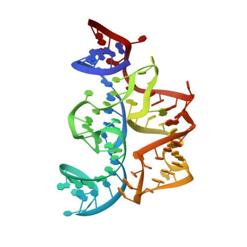
Structure of the Eukaryotic Thiamine Pyrophosphate Riboswitch with its Regulatory Ligand.

Thore, S.Leibundgut, M.Ban, N.

(2006) Science 312: 1208

  • DOI: https://doi.org/10.1126/science.1128451
  • Primary Citation of Related Structures:  
    2CKY

  • PubMed Abstract: 

    Riboswitches are untranslated regions of messenger RNA, which adopt alternate structures depending on the binding of specific metabolites. Such conformational switching regulates the expression of proteins involved in the biosynthesis of riboswitch substrates. Here, we present the 2.9 angstrom-resolution crystal structure of the eukaryotic Arabidopsis thaliana thiamine pyrophosphate (TPP)-specific riboswitch in complex with its natural ligand. The riboswitch specifically recognizes the TPP via conserved residues located within two highly distorted parallel "sensor" helices. The structure provides the basis for understanding the reorganization of the riboswitch fold upon TPP binding and explains the mechanism of resistance to the antibiotic pyrithiamine.


  • Organizational Affiliation

    ETH Zurich, Institute of Molecular Biology and Biophysics, 8092 Zurich, Switzerland. ban@mol.biol.ethz.ch


Macromolecules
Find similar nucleic acids by:  (by identity cutoff)  |  3D Structure
Entity ID: 1
MoleculeChains LengthOrganismImage
NUCLEIC ACID
A, B
77Arabidopsis thaliana
Sequence Annotations
Expand
  • Reference Sequence
Small Molecules
Ligands 3 Unique
IDChains Name / Formula / InChI Key2D Diagram3D Interactions
TPP
Query on TPP

Download Ideal Coordinates CCD File 
I [auth A],
R [auth B]
THIAMINE DIPHOSPHATE
C12 H19 N4 O7 P2 S
AYEKOFBPNLCAJY-UHFFFAOYSA-O
OS
Query on OS

Download Ideal Coordinates CCD File 
H [auth A]
J [auth A]
K [auth A]
L [auth A]
M [auth A]
H [auth A],
J [auth A],
K [auth A],
L [auth A],
M [auth A],
Q [auth B],
U [auth B],
V [auth B],
W [auth B],
X [auth B],
Y [auth B],
Z [auth B]
OSMIUM ION
Os
XQBKHDFIPARBOX-UHFFFAOYSA-N
MG
Query on MG

Download Ideal Coordinates CCD File 
C [auth A]
D [auth A]
E [auth A]
F [auth A]
G [auth A]
C [auth A],
D [auth A],
E [auth A],
F [auth A],
G [auth A],
N [auth B],
O [auth B],
P [auth B],
S [auth B],
T [auth B]
MAGNESIUM ION
Mg
JLVVSXFLKOJNIY-UHFFFAOYSA-N
Experimental Data & Validation

Experimental Data

  • Method: X-RAY DIFFRACTION
  • Resolution: 2.90 Å
  • R-Value Free: 0.250 
  • R-Value Work: 0.183 
  • R-Value Observed: 0.191 
  • Space Group: P 1 21 1
Unit Cell:
Length ( Å )Angle ( ˚ )
a = 46.85α = 90
b = 98.67β = 104.8
c = 54.55γ = 90
Software Package:
Software NamePurpose
XDSdata reduction
XDSdata scaling
SHELXphasing
SHARPphasing
REFMACrefinement

Structure Validation

View Full Validation Report


Ligand Structure Quality Assessment 


Entry History 

Deposition Data

Revision History  (Full details and data files)

  • Version 1.0: 2006-05-08
    Type: Initial release
  • Version 1.1: 2011-07-13
    Changes: Advisory, Version format compliance