2CII

The crystal structure of H-2Db complexed with a partial peptide epitope suggests an MHC Class I assembly-intermediate


Experimental Data Snapshot

  • Method: X-RAY DIFFRACTION
  • Resolution: 2.55 Å
  • R-Value Free: 0.297 
  • R-Value Work: 0.239 
  • R-Value Observed: 0.239 

wwPDB Validation   3D Report Full Report


This is version 1.4 of the entry. See complete history


Literature

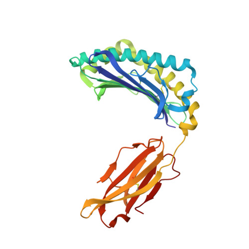
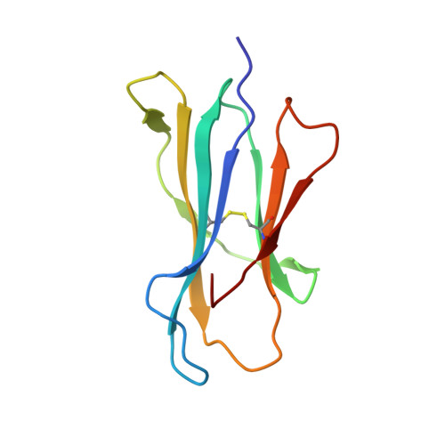
The crystal structure of H-2D(b) complexed with a partial peptide epitope suggests a major histocompatibility complex class I assembly intermediate.

Glithero, A.Tormo, J.Doering, K.Kojima, M.Jones, E.Y.Elliott, T.

(2006) J Biol Chem 281: 12699-12704

  • DOI: https://doi.org/10.1074/jbc.M511683200
  • Primary Citation of Related Structures:  
    2CII

  • PubMed Abstract: 

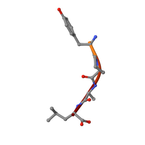
    In the absence of bound peptide ligands, major histocompatibility complex (MHC) class I molecules are unstable. In an attempt to determine the minimum requirement for peptide-dependent MHC class I stabilization, we have used short synthetic peptides derived from the Sendai virus nucleoprotein epitope (residues 324-332, 1FAPGNYPAL9) to promote its folding in vitro of H-2D(b). We found that H-2D(b) can be stabilized by the pentapeptide 5NYPAL9, which is equivalent to the C-terminal portion of the optimal nonapeptide and includes both the P5 and P9 anchor residues. We have crystallized the complex of the H-2D(b) molecule with the pentamer and determined the structure to show how a quasi-stable MHC class I molecule can be formed by occupancy of a single binding pocket in the peptide-binding groove.


  • Organizational Affiliation

    Nuffield Department of Clinical Medicine, University of Oxford, John Radcliffe Hospital, Oxford OX3 4RU, United Kingdom.


Macromolecules
Find similar proteins by:  (by identity cutoff)  |  3D Structure
Entity ID: 1
MoleculeChains Sequence LengthOrganismDetailsImage
H-2 CLASS I HISTOCOMPATIBILITY ANTIGEN D-B ALPHA CHAIN275Mus musculusMutation(s): 0 
UniProt
Find proteins for P01899 (Mus musculus)
Explore P01899 
Go to UniProtKB:  P01899
Entity Groups  
Sequence Clusters30% Identity50% Identity70% Identity90% Identity95% Identity100% Identity
UniProt GroupP01899
Sequence Annotations
Expand
  • Reference Sequence
Find similar proteins by:  (by identity cutoff)  |  3D Structure
Entity ID: 2
MoleculeChains Sequence LengthOrganismDetailsImage
BETA-2-MICROGLOBULIN99Homo sapiensMutation(s): 0 
UniProt & NIH Common Fund Data Resources
Find proteins for P61769 (Homo sapiens)
Explore P61769 
Go to UniProtKB:  P61769
PHAROS:  P61769
GTEx:  ENSG00000166710 
Entity Groups  
Sequence Clusters30% Identity50% Identity70% Identity90% Identity95% Identity100% Identity
UniProt GroupP61769
Sequence Annotations
Expand
  • Reference Sequence

Find similar proteins by:  Sequence   |   3D Structure  

Entity ID: 3
MoleculeChains Sequence LengthOrganismDetailsImage
NUCLEOPROTEIN9Human respirovirus 1Mutation(s): 0 
UniProt
Find proteins for P26590 (Human parainfluenza 1 virus (strain Washington/1957))
Explore P26590 
Go to UniProtKB:  P26590
Entity Groups  
UniProt GroupP26590
Sequence Annotations
Expand
  • Reference Sequence
Experimental Data & Validation

Experimental Data

  • Method: X-RAY DIFFRACTION
  • Resolution: 2.55 Å
  • R-Value Free: 0.297 
  • R-Value Work: 0.239 
  • R-Value Observed: 0.239 
  • Space Group: P 1 21 1
Unit Cell:
Length ( Å )Angle ( ˚ )
a = 60.996α = 90
b = 58.45β = 106.77
c = 73.534γ = 90
Software Package:
Software NamePurpose
DENZOdata reduction
SCALEPACKdata scaling
X-PLORphasing
CNSphasing
CNSrefinement

Structure Validation

View Full Validation Report


Entry History 

Deposition Data

Revision History  (Full details and data files)

  • Version 1.0: 2006-03-29
    Type: Initial release
  • Version 1.1: 2011-05-08
    Changes: Version format compliance
  • Version 1.2: 2011-07-13
    Changes: Version format compliance
  • Version 1.3: 2018-01-31
    Changes: Database references, Source and taxonomy
  • Version 1.4: 2023-12-13
    Changes: Data collection, Database references, Other, Refinement description