2C5O

Differential Binding Of Inhibitors To Active And Inactive Cdk2 Provides Insights For Drug Design


Experimental Data Snapshot

  • Method: X-RAY DIFFRACTION
  • Resolution: 2.10 Å
  • R-Value Free: 0.256 
  • R-Value Work: 0.195 
  • R-Value Observed: 0.197 

wwPDB Validation   3D Report Full Report


Ligand Structure Quality Assessment 


This is version 1.3 of the entry. See complete history


Literature

Differential Binding of Inhibitors to Active and Inactive Cdk2 Provides Insights for Drug Design.

Kontopidis, G.Mcinnes, C.Pandalaneni, S.R.Mcnae, I.Gibson, D.Mezna, M.Thomas, M.Wood, G.Wang, S.Walkinshaw, M.D.Fischer, P.M.

(2006) Chem Biol 13: 201

  • DOI: https://doi.org/10.1016/j.chembiol.2005.11.011
  • Primary Citation of Related Structures:  
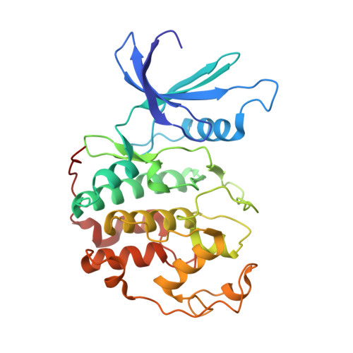
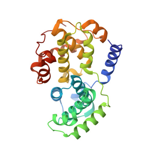
    2C5N, 2C5O, 2C5V, 2C5X, 2C5Y

  • PubMed Abstract: 

    The cyclin-dependent kinases (CDKs) have been characterized in complex with a variety of inhibitors, but the majority of structures solved are in the inactive form. We have solved the structures of six inhibitors in both the monomeric CDK2 and binary CDK2/cyclinA complexes and demonstrate that significant differences in ligand binding occur depending on the activation state. The binding mode of two ligands in particular varies substantially in active and inactive CDK2. Furthermore, energetic analysis of CDK2/cyclin/inhibitors demonstrates that a good correlation exists between the in vitro potency and the calculated energies of interaction, but no such relationship exists for CDK2/inhibitor structures. These results confirm that monomeric CDK2 ligand complexes do not fully reflect active conformations, revealing significant implications for inhibitor design while also suggesting that the monomeric CDK2 conformation can be selectively inhibited.


  • Organizational Affiliation

    Cyclacel Ltd., Dundee, UK.


Macromolecules
Find similar proteins by:  (by identity cutoff)  |  3D Structure
Entity ID: 1
MoleculeChains Sequence LengthOrganismDetailsImage
CELL DIVISION PROTEIN KINASE 2
A, C
298Homo sapiensMutation(s): 0 
EC: 2.7.1.37
UniProt & NIH Common Fund Data Resources
Find proteins for P24941 (Homo sapiens)
Explore P24941 
Go to UniProtKB:  P24941
PHAROS:  P24941
GTEx:  ENSG00000123374 
Entity Groups  
Sequence Clusters30% Identity50% Identity70% Identity90% Identity95% Identity100% Identity
UniProt GroupP24941
Sequence Annotations
Expand
  • Reference Sequence
Find similar proteins by:  (by identity cutoff)  |  3D Structure
Entity ID: 2
MoleculeChains Sequence LengthOrganismDetailsImage
CYCLIN A2
B, D
260Homo sapiensMutation(s): 0 
UniProt & NIH Common Fund Data Resources
Find proteins for P20248 (Homo sapiens)
Explore P20248 
Go to UniProtKB:  P20248
PHAROS:  P20248
GTEx:  ENSG00000145386 
Entity Groups  
Sequence Clusters30% Identity50% Identity70% Identity90% Identity95% Identity100% Identity
UniProt GroupP20248
Sequence Annotations
Expand
  • Reference Sequence
Small Molecules
Ligands 1 Unique
IDChains Name / Formula / InChI Key2D Diagram3D Interactions
CK2
Query on CK2

Download Ideal Coordinates CCD File 
E [auth A],
F [auth C]
4-(2,4-DIMETHYL-1,3-THIAZOL-5-YL)PYRIMIDIN-2-AMINE
C9 H10 N4 S
CTFDMGIBHFQWKB-UHFFFAOYSA-N
Binding Affinity Annotations 
IDSourceBinding Affinity
CK2 BindingDB:  2C5O Ki: min: 396, max: 6500 (nM) from 2 assay(s)
Kd: 3.03e+4 (nM) from 1 assay(s)
IC50: min: 6457, max: 6500 (nM) from 2 assay(s)
Binding MOAD:  2C5O IC50: 6500 (nM) from 1 assay(s)
PDBBind:  2C5O IC50: 6500 (nM) from 1 assay(s)
Experimental Data & Validation

Experimental Data

  • Method: X-RAY DIFFRACTION
  • Resolution: 2.10 Å
  • R-Value Free: 0.256 
  • R-Value Work: 0.195 
  • R-Value Observed: 0.197 
  • Space Group: P 21 21 21
Unit Cell:
Length ( Å )Angle ( ˚ )
a = 74.482α = 90
b = 113.508β = 90
c = 157.084γ = 90
Software Package:
Software NamePurpose
REFMACrefinement
DENZOdata reduction
SCALAdata scaling
MOLREPphasing

Structure Validation

View Full Validation Report


Ligand Structure Quality Assessment 


Entry History 

Deposition Data

Revision History  (Full details and data files)

  • Version 1.0: 2006-03-01
    Type: Initial release
  • Version 1.1: 2011-05-08
    Changes: Version format compliance
  • Version 1.2: 2011-07-13
    Changes: Version format compliance
  • Version 1.3: 2023-12-13
    Changes: Data collection, Database references, Other, Refinement description