2BRK

Crystal structure of Hepatitis C virus polymerase in complex with an allosteric inhibitor (compound 1)


Experimental Data Snapshot

  • Method: X-RAY DIFFRACTION
  • Resolution: 2.30 Å
  • R-Value Free: 0.245 
  • R-Value Work: 0.184 
  • R-Value Observed: 0.187 

wwPDB Validation   3D Report Full Report


Ligand Structure Quality Assessment 


This is version 1.3 of the entry. See complete history


Literature

Interdomain Communication in Hepatitis C Virus Polymerase Abolished by Small-Molecule Inhibitors Bound to a Novel Allosteric Site

Di Marco, S.Volpari, C.Tomei, L.Altamura, S.Harper, S.Narjes, F.Koch, U.Rowley, M.De Francesco, R.Migliaccio, G.Carfi, A.

(2005) J Biol Chem 280: 29765

  • DOI: https://doi.org/10.1074/jbc.M505423200
  • Primary Citation of Related Structures:  
    2BRK, 2BRL

  • PubMed Abstract: 

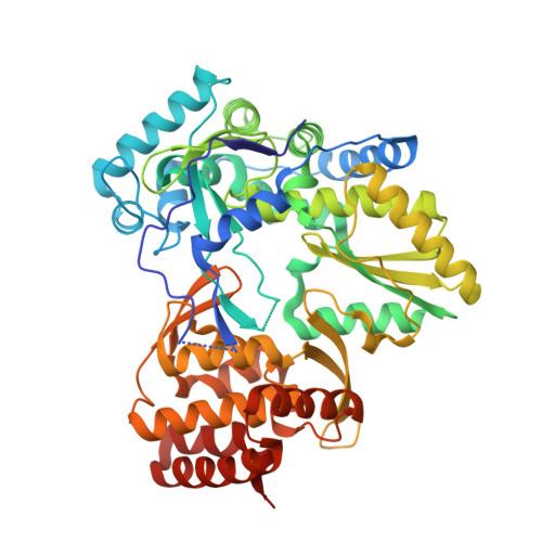
    The hepatitis C virus (HCV) polymerase is required for replication of the viral genome and is a key target for therapeutic intervention against HCV. We have determined the crystal structures of the HCV polymerase complexed with two indole-based allosteric inhibitors at 2.3- and 2.4-Angstroms resolution. The structures show that these inhibitors bind to a site on the surface of the thumb domain. A cyclohexyl and phenyl ring substituents, bridged by an indole moiety, fill two closely spaced pockets, whereas a carboxylate substituent forms a salt bridge with an exposed arginine side chain. Interestingly, in the apoenzyme, the inhibitor binding site is occupied by a small alpha-helix at the tip of the N-terminal loop that connects the fingers and thumb domains. Thus, these molecules inhibit the enzyme by preventing formation of intramolecular contacts between these two domains and consequently precluding their coordinated movements during RNA synthesis. Our structures identify a novel mechanism by which a new class of allosteric inhibitors inhibits the HCV polymerase and open the way to the development of novel antiviral agents against this clinically relevant human pathogen.


  • Organizational Affiliation

    Istituto di Ricerche di Biologia Molecolare P. Angeletti, Pomezia (Rome), Italy.


Macromolecules
Find similar proteins by:  (by identity cutoff)  |  3D Structure
Entity ID: 1
MoleculeChains Sequence LengthOrganismDetailsImage
RNA-DIRECTED RNA POLYMERASE536Hepacivirus hominisMutation(s): 0 
EC: 2.7.7.48
UniProt
Find proteins for P26663 (Hepatitis C virus genotype 1b (isolate BK))
Explore P26663 
Go to UniProtKB:  P26663
Entity Groups  
Sequence Clusters30% Identity50% Identity70% Identity90% Identity95% Identity100% Identity
UniProt GroupP26663
Sequence Annotations
Expand
  • Reference Sequence
Small Molecules
Ligands 2 Unique
IDChains Name / Formula / InChI Key2D Diagram3D Interactions
CMF
Query on CMF

Download Ideal Coordinates CCD File 
D [auth A]3-CYCLOHEXYL-1-(2-MORPHOLIN-4-YL-2-OXOETHYL)-2-PHENYL-1H-INDOLE-6-CARBOXYLIC ACID
C27 H30 N2 O4
ZKEZEXYKYHYIMQ-UHFFFAOYSA-N
MN
Query on MN

Download Ideal Coordinates CCD File 
B [auth A],
C [auth A]
MANGANESE (II) ION
Mn
WAEMQWOKJMHJLA-UHFFFAOYSA-N
Experimental Data & Validation

Experimental Data

  • Method: X-RAY DIFFRACTION
  • Resolution: 2.30 Å
  • R-Value Free: 0.245 
  • R-Value Work: 0.184 
  • R-Value Observed: 0.187 
  • Space Group: P 21 21 2
Unit Cell:
Length ( Å )Angle ( ˚ )
a = 68.002α = 90
b = 94.488β = 90
c = 95.73γ = 90
Software Package:
Software NamePurpose
REFMACrefinement
MOSFLMdata reduction
SCALAdata scaling
AMoREphasing

Structure Validation

View Full Validation Report


Ligand Structure Quality Assessment 


Entry History 

Deposition Data

Revision History  (Full details and data files)

  • Version 1.0: 2005-06-14
    Type: Initial release
  • Version 1.1: 2011-05-08
    Changes: Version format compliance
  • Version 1.2: 2011-07-13
    Changes: Version format compliance
  • Version 1.3: 2023-12-13
    Changes: Data collection, Database references, Derived calculations, Other, Refinement description