2BPX

HIV-1 protease-inhibitor complex


Experimental Data Snapshot

  • Method: X-RAY DIFFRACTION
  • Resolution: 2.80 Å
  • R-Value Work: 0.175 
  • R-Value Observed: 0.175 

wwPDB Validation   3D Report Full Report


This is version 1.4 of the entry. See complete history


Literature

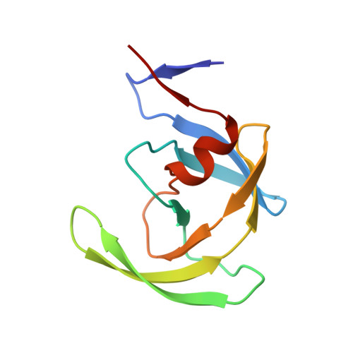
Rapid X-ray diffraction analysis of HIV-1 protease-inhibitor complexes: inhibitor exchange in single crystals of the bound enzyme.

Munshi, S.Chen, Z.Li, Y.Olsen, D.B.Fraley, M.E.Hungate, R.W.Kuo, L.C.

(1998) Acta Crystallogr D Biol Crystallogr 54: 1053-1060

  • DOI: https://doi.org/10.1107/s0907444998003588
  • Primary Citation of Related Structures:  
    2BPV, 2BPW, 2BPX, 2BPY, 2BPZ

  • PubMed Abstract: 

    The ability to replace an inhibitor bound to the HIV-1 protease in single crystals with other potent inhibitors offers the possibility of investigating a series of protease inhibitors rapidly and conveniently with the use of X-ray crystallography. This approach affords a fast turnaround of structural information for iterative rational drug designs and obviates the need for studying the complex structures by co-crystallization. The replacement approach has been successfully used with single crystals of the HIV-1 protease complexed with a weak inhibitor. The structures of the complexes obtained by the replacement method are similar to those determined by co-crystallization.


  • Organizational Affiliation

    Department of Antiviral Research, Merck Research Laboratories, West Point, PA 19486, USA.


Macromolecules
Find similar proteins by:  (by identity cutoff)  |  3D Structure
Entity ID: 1
MoleculeChains Sequence LengthOrganismDetailsImage
HIV-1 PROTEASE
A, B
99Human immunodeficiency virus 1Mutation(s): 0 
Gene Names: HIV-1 PROTEASE FROM THE NY5 ISOLATE
EC: 3.4.23.16
UniProt
Find proteins for P04587 (Human immunodeficiency virus type 1 group M subtype B (isolate BH5))
Explore P04587 
Go to UniProtKB:  P04587
Entity Groups  
Sequence Clusters30% Identity50% Identity70% Identity90% Identity95% Identity100% Identity
UniProt GroupP04587
Sequence Annotations
Expand
  • Reference Sequence
Small Molecules
Ligands 1 Unique
IDChains Name / Formula / InChI Key2D Diagram3D Interactions
MK1
Query on MK1

Download Ideal Coordinates CCD File 
C [auth B]N-[2(R)-HYDROXY-1(S)-INDANYL]-5-[(2(S)-TERTIARY BUTYLAMINOCARBONYL)-4(3-PYRIDYLMETHYL)PIPERAZINO]-4(S)-HYDROXY-2(R)-PHENYLMETHYLPENTANAMIDE
C36 H47 N5 O4
CBVCZFGXHXORBI-PXQQMZJSSA-N
Binding Affinity Annotations 
IDSourceBinding Affinity
MK1 BindingDB:  2BPX Ki: min: 0.05, max: 398 (nM) from 50 assay(s)
Kd: min: 1.07, max: 5.5 (nM) from 4 assay(s)
IC50: min: 0.36, max: 370 (nM) from 23 assay(s)
-TΔS: min: -8.28e+1, max: -2.80e+1 (kJ/mol) from 38 assay(s)
ΔH: min: -3.34e+1, max: 51 (kJ/mol) from 39 assay(s)
ΔG: min: -6.31e+1, max: -3.18e+1 (kJ/mol) from 37 assay(s)
PDBBind:  2BPX Ki: 0.4 (nM) from 1 assay(s)
Experimental Data & Validation

Experimental Data

  • Method: X-RAY DIFFRACTION
  • Resolution: 2.80 Å
  • R-Value Work: 0.175 
  • R-Value Observed: 0.175 
  • Space Group: P 21 21 2
Unit Cell:
Length ( Å )Angle ( ˚ )
a = 58.6α = 90
b = 87.37β = 90
c = 46.64γ = 90
Software Package:
Software NamePurpose
X-PLORmodel building
X-PLORrefinement
X-PLORphasing

Structure Validation

View Full Validation Report


Entry History 

Deposition Data

Revision History  (Full details and data files)

  • Version 1.0: 1999-02-23
    Type: Initial release
  • Version 1.1: 2008-03-24
    Changes: Version format compliance
  • Version 1.2: 2011-07-13
    Changes: Version format compliance
  • Version 1.3: 2012-05-09
    Changes: Structure summary
  • Version 1.4: 2024-02-14
    Changes: Data collection, Database references, Derived calculations, Other