2BP3

Crystal structure of Filamin A domain 17 and GPIb alpha cytoplasmic domain complex


Experimental Data Snapshot

  • Method: X-RAY DIFFRACTION
  • Resolution: 2.32 Å
  • R-Value Free: 0.256 
  • R-Value Work: 0.214 
  • R-Value Observed: 0.216 

wwPDB Validation   3D Report Full Report


This is version 1.3 of the entry. See complete history


Literature

The Structure of the Gpib-Filamin a Complex.

Nakamura, F.Pudas, R.Heikkinen, O.Permi, P.Kilpelainen, I.Munday, A.D.Hartwig, J.H.Stossel, T.P.Ylanne, J.

(2006) Blood 107: 1925

  • DOI: https://doi.org/10.1182/blood-2005-10-3964
  • Primary Citation of Related Structures:  
    2AAV, 2BP3

  • PubMed Abstract: 

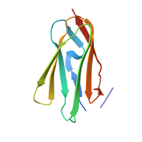
    Filamin A (FLNa), a dimeric actin cross-linking and scaffold protein with numerous intracellular binding partners, anchors the platelet adhesion glycoprotein (GP) Ib-IX-V receptor to actin cytoskeleton. We mapped the GPIbalpha binding site to a single domain of FLNa and resolved the structure of this domain and its interaction complex with the corresponding GPIbalpha cytoplasmic domain. This is the first atomic structure of this class of membrane glycoprotein-cytoskeleton connection. GPIbalpha binds in a groove formed between the C and D beta strands of FLNa domain 17. The interaction is strikingly similar to that between the beta7 integrin tail and a different FLNa domain, potentially defining a conserved motif for FLNa binding. Nevertheless, the structures also reveal specificity of the interfaces, which explains different regulatory mechanisms. To verify the topology of GPIb-FLNa interaction we also purified the native complex from platelets and showed that GPIb interacts with the C-terminus of FLNa, which is in accordance with our biochemical and structural data.


  • Organizational Affiliation

    Hematology Division, Department of Medicine, Brigham and Women's Hospital, Harvard Medical School, Boston, MA, USA.


Macromolecules
Find similar proteins by:  (by identity cutoff)  |  3D Structure
Entity ID: 1
MoleculeChains Sequence LengthOrganismDetailsImage
FILAMIN A
A, B
97Homo sapiensMutation(s): 0 
UniProt & NIH Common Fund Data Resources
Find proteins for P21333 (Homo sapiens)
Explore P21333 
Go to UniProtKB:  P21333
PHAROS:  P21333
GTEx:  ENSG00000196924 
Entity Groups  
Sequence Clusters30% Identity50% Identity70% Identity90% Identity95% Identity100% Identity
UniProt GroupP21333
Sequence Annotations
Expand
  • Reference Sequence

Find similar proteins by:  Sequence   |   3D Structure  

Entity ID: 2
MoleculeChains Sequence LengthOrganismDetailsImage
PLATELET GLYCOPROTEIN IB ALPHA CHAINC [auth S],
D [auth T]
22Homo sapiensMutation(s): 0 
UniProt & NIH Common Fund Data Resources
Find proteins for P07359 (Homo sapiens)
Explore P07359 
Go to UniProtKB:  P07359
PHAROS:  P07359
GTEx:  ENSG00000185245 
Entity Groups  
Sequence Clusters30% Identity50% Identity70% Identity90% Identity95% Identity100% Identity
UniProt GroupP07359
Sequence Annotations
Expand
  • Reference Sequence
Experimental Data & Validation

Experimental Data

  • Method: X-RAY DIFFRACTION
  • Resolution: 2.32 Å
  • R-Value Free: 0.256 
  • R-Value Work: 0.214 
  • R-Value Observed: 0.216 
  • Space Group: P 21 21 21
Unit Cell:
Length ( Å )Angle ( ˚ )
a = 41.9α = 90
b = 62.8β = 90
c = 122.8γ = 90
Software Package:
Software NamePurpose
REFMACrefinement
XDSdata reduction
XDSdata scaling
PHASERphasing

Structure Validation

View Full Validation Report


Entry History 

Deposition Data

Revision History  (Full details and data files)

  • Version 1.0: 2005-11-30
    Type: Initial release
  • Version 1.1: 2011-05-08
    Changes: Version format compliance
  • Version 1.2: 2011-07-13
    Changes: Version format compliance
  • Version 1.3: 2023-12-13
    Changes: Data collection, Database references, Other, Refinement description