2BGD

Structure-based design of Protein Tyrosine Phosphatase-1B Inhibitors


Experimental Data Snapshot

  • Method: X-RAY DIFFRACTION
  • Resolution: 2.40 Å
  • R-Value Free: 0.200 
  • R-Value Work: 0.164 
  • R-Value Observed: 0.166 

wwPDB Validation   3D Report Full Report


Ligand Structure Quality Assessment 


This is version 1.3 of the entry. See complete history


Literature

Structure-Based Design of Protein Tyrosine Phosphatase-1B Inhibitors

Black, E.Breed, J.Breeze, A.L.Embrey, K.Garcia, R.Gero, T.W.Godfrey, L.Kenny, P.W.Morley, A.D.Minshull, C.A.Pannifer, A.D.Read, J.Rees, A.Russell, D.J.Toader, D.Tucker, J.

(2005) Bioorg Med Chem Lett 15: 2503

  • DOI: https://doi.org/10.1016/j.bmcl.2005.03.068
  • Primary Citation of Related Structures:  
    2BGD, 2BGE

  • PubMed Abstract: 

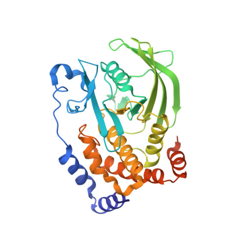
    Using structure-based design, a new class of inhibitors of protein tyrosine phosphatase-1B (PTP1B) has been identified, which incorporate the 1,2,5-thiadiazolidin-3-one-1,1-dioxide template.


  • Organizational Affiliation

    AstraZeneca, Mereside, Alderley Park, Macclesfield, Cheshire SK10 4TG, UK.


Macromolecules
Find similar proteins by:  (by identity cutoff)  |  3D Structure
Entity ID: 1
MoleculeChains Sequence LengthOrganismDetailsImage
PROTEIN-TYROSINE PHOSPHATASE NON-RECEPTOR TYPE 1321Homo sapiensMutation(s): 0 
EC: 3.1.3.48
UniProt & NIH Common Fund Data Resources
Find proteins for P18031 (Homo sapiens)
Explore P18031 
Go to UniProtKB:  P18031
PHAROS:  P18031
GTEx:  ENSG00000196396 
Entity Groups  
Sequence Clusters30% Identity50% Identity70% Identity90% Identity95% Identity100% Identity
UniProt GroupP18031
Sequence Annotations
Expand
  • Reference Sequence
Small Molecules
Binding Affinity Annotations 
IDSourceBinding Affinity
T1D BindingDB:  2BGD Ki: 2400 (nM) from 1 assay(s)
IC50: min: 2470, max: 4800 (nM) from 3 assay(s)
PDBBind:  2BGD Kd: 1.00e+4 (nM) from 1 assay(s)
Binding MOAD:  2BGD IC50: 2470 (nM) from 1 assay(s)
Experimental Data & Validation

Experimental Data

  • Method: X-RAY DIFFRACTION
  • Resolution: 2.40 Å
  • R-Value Free: 0.200 
  • R-Value Work: 0.164 
  • R-Value Observed: 0.166 
  • Space Group: P 31 2 1
Unit Cell:
Length ( Å )Angle ( ˚ )
a = 88.908α = 90
b = 88.908β = 90
c = 104.548γ = 120
Software Package:
Software NamePurpose
REFMACrefinement
MOSFLMdata reduction
SCALAdata scaling
AMoREphasing

Structure Validation

View Full Validation Report


Ligand Structure Quality Assessment 


Entry History 

Revision History  (Full details and data files)

  • Version 1.0: 2005-05-04
    Type: Initial release
  • Version 1.1: 2011-07-13
    Changes: Advisory, Refinement description, Version format compliance
  • Version 1.2: 2015-09-16
    Changes: Source and taxonomy
  • Version 1.3: 2019-07-24
    Changes: Data collection