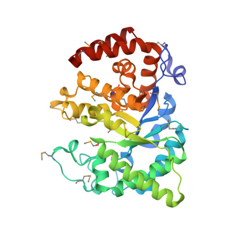
Crystal Structure of the Phosphoenolpyruvate-Binding Enzyme I-Domain from the Thermoanaerobacter Tengcongensis Pep: Sugar Phosphotransferase System (Pts)
Oberholzer, A.E., Bumann, M., Schneider, P., Baechler, C., Siebold, C., Baumann, U., Erni, B.(2005) J Mol Biol 346: 521
- PubMed: 15670601
- DOI: https://doi.org/10.1016/j.jmb.2004.11.077
- Primary Citation of Related Structures:
2BG5 - PubMed Abstract:
Enzyme I (EI), the first component of the phosphoenolpyruvate (PEP):sugar phosphotransferase system (PTS), consists of an N-terminal protein-binding domain (EIN) and a C-terminal PEP-binding domain (EIC). EI transfers phosphate from PEP by double displacement via a histidine residue on EIN to the general phosphoryl carrier protein HPr. Here, we report the 1.82A crystal structure of the homodimeric EIC domain from Thermoanaerobacter tengcongensis, a saccharolytic eubacterium that grows optimally at 75 degrees C. EIC folds into a (betaalpha)(8) barrel with three large helical insertions between beta2/alpha2, beta3/alpha3 and beta6/alpha6. The large amphipathic dimer interface buries 3750A(2) of accessible surface area per monomer. A comparison with pyruvate phosphate dikinase (PPDK) reveals that the active-site residues in the empty PEP-binding site of EIC and in the liganded PEP-binding site of PPDK have almost identical conformations, pointing to a rigid structure of the active site. In silico models of EIC in complex with the Z and E-isomers of chloro-PEP provide a rational explanation for their difference as substrates and inhibitors of EI. The EIC domain exhibits 54% amino acid sequence identity with Escherichia coli and 60% with Bacillus subtilis EIC, has the same amino acid composition but contains additional salt-bridges and a more complex salt-bridge network than the homology model of E.coli EIC. The easy crystallization of EIC suggests that T.tengcongensis can serve as source for stable homologs of mesophilic proteins that are too labile for crystallization.
Organizational Affiliation:
Department of Chemistry and Biochemistry, University of Berne, Freiestrasse 3, CH-3012 Bern, Switzerland.