Competitive Inhibitors of Mycobacterium Tuberculosis Ribose-5-Phosphate Isomerase B Reveal New Information About the Reaction Mechanism
Roos, A.K., Burgos, E., Ericsson, D.J., Salmon, L., Mowbray, S.L.(2005) J Biol Chem 280: 6416
- PubMed: 15590681
- DOI: https://doi.org/10.1074/jbc.M412018200
- Primary Citation of Related Structures:
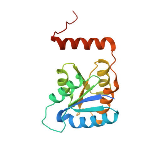
2BES, 2BET - PubMed Abstract:
Ribose-5-phosphate isomerase (Rpi), an important enzyme in the pentose phosphate pathway, catalyzes the interconversion of ribulose 5-phosphate and ribose 5-phosphate. Two unrelated isomerases have been identified, RpiA and RpiB, with different structures and active site residues. The reaction catalyzed by both enzymes is thought to proceed via a high energy enediolate intermediate, by analogy to other carbohydrate isomerases. Here we present studies of RpiB from Mycobacterium tuberculosis together with small molecules designed to resemble the enediolate intermediate. The relative affinities of these inhibitors for RpiB have a different pattern than that observed previously for the RpiA from spinach. X-ray structures of RpiB in complex with the inhibitors 4-phospho-d-erythronohydroxamic acid (K(m) 57 microm) and 4-phospho-d-erythronate (K(i) 1.7 mm) refined to resolutions of 2.1 and 2.2 A, respectively, allowed us to assign roles for most active site residues. These results, combined with docking of the substrates in the position of the most effective inhibitor, now allow us to outline the reaction mechanism for RpiBs. Both enzymes have residues that can catalyze opening of the furanose ring of the ribose 5-phosphate and so can improve the efficiency of the reaction. Both enzymes also have an acidic residue that acts as a base in the isomerization step. A lysine residue in RpiAs provides for more efficient stabilization of the intermediate than the corresponding uncharged groups of RpiBs; this same feature lies behind the more efficient binding of RpiA to 4-phospho-d-erythronate.
Organizational Affiliation:
Department of Cell and Molecular Biology, Uppsala University, Biomedical Center, Box 596, Uppsala SE-751 24, Sweden.