2BDN

Crystal structure of human MCP-1 bound to a blocking antibody, 11K2


Experimental Data Snapshot

  • Method: X-RAY DIFFRACTION
  • Resolution: 2.53 Å
  • R-Value Free: 0.277 
  • R-Value Work: 0.219 
  • R-Value Observed: 0.220 

wwPDB Validation   3D Report Full Report


This is version 1.4 of the entry. See complete history


Literature

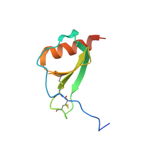
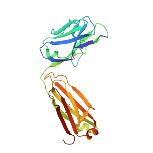
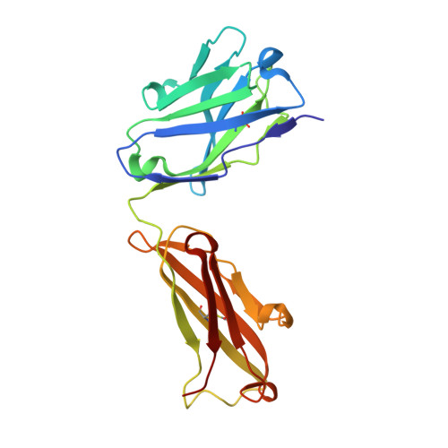
Structure activity relationships of monocyte chemoattractant proteins in complex with a blocking antibody.

Reid, C.Rushe, M.Jarpe, M.van Vlijmen, H.Dolinski, B.Qian, F.Cachero, T.G.Cuervo, H.Yanachkova, M.Nwankwo, C.Wang, X.Etienne, N.Garber, E.Bailly, V.de Fougerolles, A.Boriack-Sjodin, P.A.

(2006) Protein Eng Des Sel 19: 317-324

  • DOI: https://doi.org/10.1093/protein/gzl015
  • Primary Citation of Related Structures:  
    2BDN

  • PubMed Abstract: 

    Monocyte chemoattractant proteins (MCPs) are cytokines that direct immune cells bearing appropriate receptors to sites of inflammation or injury and are therefore attractive therapeutic targets for inhibitory molecules. 11K2 is a blocking mouse monoclonal antibody active against several human and murine MCPs. A 2.5 A structure of the Fab fragment of this antibody in complex with human MCP-1 has been solved. The Fab blocks CCR2 receptor binding to MCP-1 through an adjacent but distinct binding site. The orientation of the Fab indicates that a single MCP-1 dimer will bind two 11K2 antibodies. Several key residues on the antibody and on human MCPs were predicted to be involved in antibody selectivity. Mutational analysis of these residues confirms their involvement in the antibody-chemokine interaction. In addition to mutations that decreased or disrupted binding, one antibody mutation resulted in a 70-fold increase in affinity for human MCP-2. A key residue missing in human MCP-3, a chemokine not recognized by the antibody, was identified and engineering the preferred residue into the chemokine conferred binding to the antibody.


  • Organizational Affiliation

    Department of Research, Biogen Idec, Inc. 12 Cambridge Center, Cambridge, MA 02142, USA.


Macromolecules
Find similar proteins by:  (by identity cutoff)  |  3D Structure
Entity ID: 1
MoleculeChains Sequence LengthOrganismDetailsImage
Small inducible cytokine A276N/AMutation(s): 0 
UniProt & NIH Common Fund Data Resources
Find proteins for P13500 (Homo sapiens)
Explore P13500 
Go to UniProtKB:  P13500
PHAROS:  P13500
GTEx:  ENSG00000108691 
Entity Groups  
Sequence Clusters30% Identity50% Identity70% Identity90% Identity95% Identity100% Identity
UniProt GroupP13500
Sequence Annotations
Expand
  • Reference Sequence
Find similar proteins by:  (by identity cutoff)  |  3D Structure
Entity ID: 2
MoleculeChains Sequence LengthOrganismDetailsImage
Antibody light chain 11K2B [auth L]214Mus musculusMutation(s): 0 
Entity Groups  
Sequence Clusters30% Identity50% Identity70% Identity90% Identity95% Identity100% Identity
Sequence Annotations
Expand
  • Reference Sequence
Find similar proteins by:  (by identity cutoff)  |  3D Structure
Entity ID: 3
MoleculeChains Sequence LengthOrganismDetailsImage
Antibody heavy chain 11K2C [auth H]217Mus musculusMutation(s): 0 
Entity Groups  
Sequence Clusters30% Identity50% Identity70% Identity90% Identity95% Identity100% Identity
Sequence Annotations
Expand
  • Reference Sequence
Experimental Data & Validation

Experimental Data

  • Method: X-RAY DIFFRACTION
  • Resolution: 2.53 Å
  • R-Value Free: 0.277 
  • R-Value Work: 0.219 
  • R-Value Observed: 0.220 
  • Space Group: C 2 2 21
Unit Cell:
Length ( Å )Angle ( ˚ )
a = 86.36α = 90
b = 89.1β = 90
c = 176.24γ = 90
Software Package:
Software NamePurpose
CNSrefinement
PDB_EXTRACTdata extraction
DENZOdata reduction
SCALEPACKdata scaling
AMoREphasing
CNXrefinement

Structure Validation

View Full Validation Report


Entry History 

Deposition Data

Revision History  (Full details and data files)

  • Version 1.0: 2006-06-13
    Type: Initial release
  • Version 1.1: 2008-05-01
    Changes: Version format compliance
  • Version 1.2: 2011-07-13
    Changes: Version format compliance
  • Version 1.3: 2018-01-24
    Changes: Structure summary
  • Version 1.4: 2024-04-03
    Changes: Data collection, Database references, Refinement description