Single-stranded DNA mimicry in the p53 transactivation domain interaction with replication protein A.
Bochkareva, E., Kaustov, L., Ayed, A., Yi, G.S., Lu, Y., Pineda-Lucena, A., Liao, J.C., Okorokov, A.L., Milner, J., Arrowsmith, C.H., Bochkarev, A.(2005) Proc Natl Acad Sci U S A 102: 15412-15417
- PubMed: 16234232
- DOI: https://doi.org/10.1073/pnas.0504614102
- Primary Citation of Related Structures:
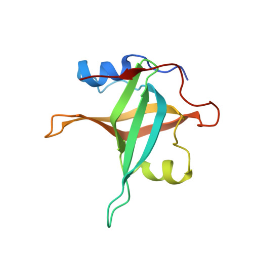
2B29, 2B3G - PubMed Abstract:
One of many protein-protein interactions modulated upon DNA damage is that of the single-stranded DNA-binding protein, replication protein A (RPA), with the p53 tumor suppressor. Here we report the crystal structure of RPA residues 1-120 (RPA70N) bound to the N-terminal transactivation domain of p53 (residues 37-57; p53N) and, by using NMR spectroscopy, characterize two mechanisms by which the RPA/p53 interaction can be modulated. RPA70N forms an oligonucleotide/oligosaccharide-binding fold, similar to that previously observed for the ssDNA-binding domains of RPA. In contrast, the N-terminal p53 transactivation domain is largely disordered in solution, but residues 37-57 fold into two amphipathic helices, H1 and H2, upon binding with RPA70N. The H2 helix of p53 structurally mimics the binding of ssDNA to the oligonucleotide/oligosaccharide-binding fold. NMR experiments confirmed that both ssDNA and an acidic peptide mimicking a phosphorylated form of RPA32N can independently compete the acidic p53N out of the binding site. Taken together, our data suggest a mechanism for DNA damage signaling that can explain a threshold response to DNA damage.
Organizational Affiliation:
Banting and Best Department of Medical Research & Department of Medical Genetics and Microbiology, University of Toronto, 112 College Street, Toronto, Ontario, Canada M5G 1L6.