2AZE

Structure of the Rb C-terminal domain bound to an E2F1-DP1 heterodimer


Experimental Data Snapshot

  • Method: X-RAY DIFFRACTION
  • Resolution: 2.55 Å
  • R-Value Free: 0.262 
  • R-Value Work: 0.221 

wwPDB Validation   3D Report Full Report


This is version 1.3 of the entry. See complete history


Literature

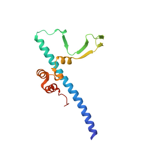
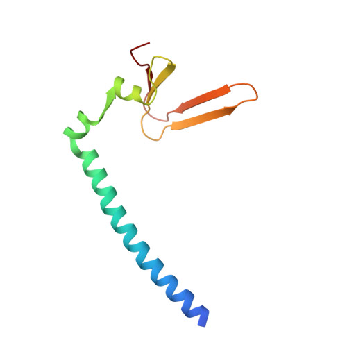
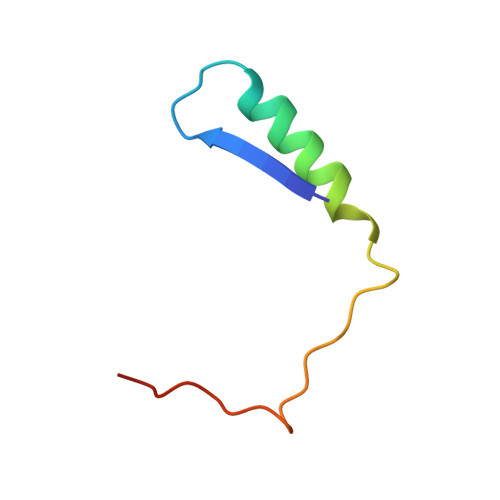
Structure of the Rb C-terminal domain bound to E2F1-DP1: a mechanism for phosphorylation-induced E2F release.

Rubin, S.M.Gall, A.L.Zheng, N.Pavletich, N.P.

(2005) Cell 123: 1093-1106

  • DOI: https://doi.org/10.1016/j.cell.2005.09.044
  • Primary Citation of Related Structures:  
    2AZE

  • PubMed Abstract: 

    The retinoblastoma (Rb) protein negatively regulates the G1-S transition by binding to the E2F transcription factors, until cyclin-dependent kinases phosphorylate Rb, causing E2F release. The Rb pocket domain is necessary for E2F binding, but the Rb C-terminal domain (RbC) is also required for growth suppression. Here we demonstrate a high-affinity interaction between RbC and E2F-DP heterodimers shared by all Rb and E2F family members. The crystal structure of an RbC-E2F1-DP1 complex reveals an intertwined heterodimer in which the marked box domains of both E2F1 and DP1 contact RbC. We also demonstrate that phosphorylation of RbC at serines 788 and 795 destabilizes one set of RbC-E2F-DP interactions directly, while phosphorylation at threonines 821 and 826 induces an intramolecular interaction between RbC and the Rb pocket that destabilizes the remaining interactions indirectly. Our findings explain the requirement of RbC for high-affinity E2F binding and growth suppression and establish a mechanism for the regulation of Rb-E2F association by phosphorylation.


  • Organizational Affiliation

    Structural Biology Program and Howard Hughes Medical Institute, Memorial Sloan-Kettering Cancer Center, NY 10021, USA.


Macromolecules
Find similar proteins by:  (by identity cutoff)  |  3D Structure
Entity ID: 1
MoleculeChains Sequence LengthOrganismDetailsImage
Transcription factor Dp-1155Homo sapiensMutation(s): 0 
Gene Names: TFDP1DP1
UniProt & NIH Common Fund Data Resources
Find proteins for Q14186 (Homo sapiens)
Explore Q14186 
Go to UniProtKB:  Q14186
PHAROS:  Q14186
GTEx:  ENSG00000198176 
Entity Groups  
Sequence Clusters30% Identity50% Identity70% Identity90% Identity95% Identity100% Identity
UniProt GroupQ14186
Sequence Annotations
Expand
  • Reference Sequence
Find similar proteins by:  (by identity cutoff)  |  3D Structure
Entity ID: 2
MoleculeChains Sequence LengthOrganismDetailsImage
Transcription factor E2F1106Homo sapiensMutation(s): 0 
Gene Names: E2F1RBBP3
UniProt & NIH Common Fund Data Resources
Find proteins for Q01094 (Homo sapiens)
Explore Q01094 
Go to UniProtKB:  Q01094
PHAROS:  Q01094
GTEx:  ENSG00000101412 
Entity Groups  
Sequence Clusters30% Identity50% Identity70% Identity90% Identity95% Identity100% Identity
UniProt GroupQ01094
Sequence Annotations
Expand
  • Reference Sequence
Find similar proteins by:  (by identity cutoff)  |  3D Structure
Entity ID: 3
MoleculeChains Sequence LengthOrganismDetailsImage
Retinoblastoma-associated protein46Homo sapiensMutation(s): 0 
Gene Names: RB1
UniProt & NIH Common Fund Data Resources
Find proteins for P06400 (Homo sapiens)
Explore P06400 
Go to UniProtKB:  P06400
PHAROS:  P06400
GTEx:  ENSG00000139687 
Entity Groups  
Sequence Clusters30% Identity50% Identity70% Identity90% Identity95% Identity100% Identity
UniProt GroupP06400
Sequence Annotations
Expand
  • Reference Sequence
Experimental Data & Validation

Experimental Data

  • Method: X-RAY DIFFRACTION
  • Resolution: 2.55 Å
  • R-Value Free: 0.262 
  • R-Value Work: 0.221 
  • Space Group: C 2 2 21
Unit Cell:
Length ( Å )Angle ( ˚ )
a = 146.8α = 90
b = 168.6β = 90
c = 48.3γ = 90
Software Package:
Software NamePurpose
DENZOdata reduction
SCALEPACKdata scaling
SHARPphasing
CNSrefinement

Structure Validation

View Full Validation Report


Entry History 

Deposition Data

Revision History  (Full details and data files)

  • Version 1.0: 2006-01-31
    Type: Initial release
  • Version 1.1: 2008-05-01
    Changes: Version format compliance
  • Version 1.2: 2011-07-13
    Changes: Version format compliance
  • Version 1.3: 2024-02-14
    Changes: Data collection, Database references