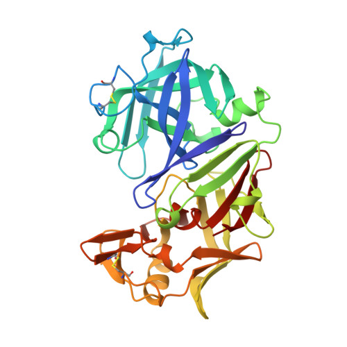
Structure and refinement at 1.8 A resolution of the aspartic proteinase from Rhizopus chinensis.
Suguna, K., Bott, R.R., Padlan, E.A., Subramanian, E., Sheriff, S., Cohen, G.H., Davies, D.R.(1987) J Mol Biol 196: 877-900
- PubMed: 3316666
- DOI: https://doi.org/10.1016/0022-2836(87)90411-6
- Primary Citation of Related Structures:
2APR - PubMed Abstract:
The structure of rhizopuspepsin (EC 3.4.23.6), the aspartic proteinase from Rhizopus chinensis, has been refined to a crystallographic R-factor of 0.143 at 1.8 A resolution. The positions of 2417 protein atoms have been determined with a root-mean-square (r.m.s.) error of 0.12 A. In the final model, the r.m.s. deviation from ideality for bond distances is 0.010 A, and for angle distances it is 0.034 A. During the course of the refinement, a calcium ion and 373 water molecules, of which 17 are internal, have been located. The active aspartate residues, Asp35 and Asp218, are involved in similar hydrogen-bonding interactions with neighboring residues and with several water molecules. One water molecule is located between the two carboxyl groups of the catalytic aspartate residues in a tightly hydrogen-bonded position. The refinement resulted in an unambiguous interpretation of the highly mobile "flap", a beta-hairpin loop region that projects over the binding pocket. Large solvent channels are formed when the molecules pack in the crystal, exposing the binding pocket and making it easily accessible. Intermolecular contacts involve mainly solvent molecules and a few protein atoms. The three-dimensional structure of rhizopuspepsin closely resembles other aspartic proteinase structures. A detailed comparison with the structure of penicillopepsin showed striking similarities as well as subtle differences in the active site geometry and molecular packing.
Organizational Affiliation:
Laboratory of Molecular Biology, National Institute of Diabetes and Digestive and Kidney Diseases, Bethesda, MD 20892.