2ADF

Crystal Structure and Paratope Determination of 82D6A3, an Antithrombotic Antibody Directed Against the von Willebrand factor A3-Domain


Experimental Data Snapshot

  • Method: X-RAY DIFFRACTION
  • Resolution: 1.90 Å
  • R-Value Free: 0.220 
  • R-Value Work: 0.191 
  • R-Value Observed: 0.192 

wwPDB Validation   3D Report Full Report


This is version 1.3 of the entry. See complete history


Literature

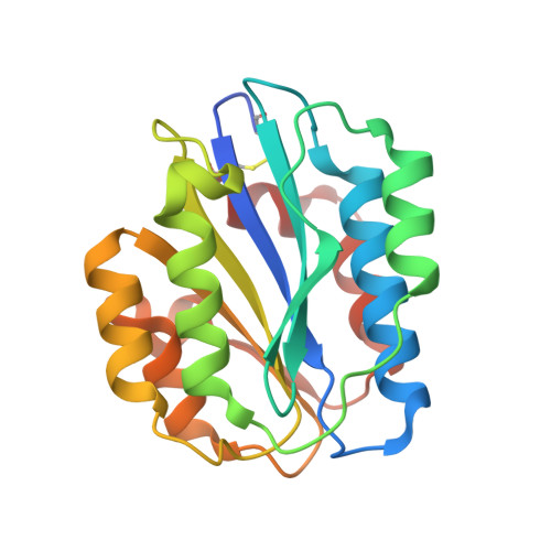
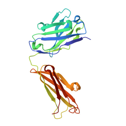
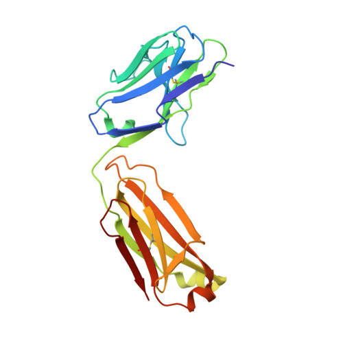
Paratope determination of the antithrombotic antibody 82D6A3 based on the crystal structure of its complex with the von Willebrand factor A3-domain

Staelens, S.Hadders, M.A.Vauterin, S.Platteau, C.De Maeyer, M.Vanhoorelbeke, K.Huizinga, E.G.Deckmyn, H.

(2006) J Biol Chem 281: 2225-2231

  • DOI: https://doi.org/10.1074/jbc.M508191200
  • Primary Citation of Related Structures:  
    2ADF

  • PubMed Abstract: 

    The antithrombotic monoclonal antibody 82D6A3 is directed against amino acids Arg-963, Pro-981, Asp-1009, Arg-1016, Ser-1020, Met-1022, and His-1023 of the von Willebrand factor A3-domain (Vanhoorelbeke, K., Depraetere, H., Romijn, R. A., Huizinga, E., De Maeyer, M., and Deckmyn, H. (2003) J. Biol. Chem. 278, 37815-37821). By this, it potently inhibits the interaction of von Willebrand factor to collagens, which is a prerequisite for blood platelet adhesion to the injured vessel wall at sites of high shear. To fully understand the mode of action of 82D6A3 at the molecular level, we resolved its crystal structure in complex with the A3-domain and fine mapped its paratope by construction and characterization of 13 mutants. The paratope predominantly consists of two short sequences in the heavy chain CDR1 (Asn-31 and Tyr-32) and CDR3 (Asp-99, Pro-101, Tyr-102 and Tyr-103), forming one patch on the surface of the antibody. Trp-50 of the heavy and His-49 of the light chain, both situated adjacent to the patch, play ancillary roles in antigen binding. The crystal structure furthermore confirms the epitope location, which largely overlaps with the collagen binding site deduced from mutagenesis of the A3-domain (Romijn, R. A., Westein, E., Bouma, B., Schiphorst, M. E., Sixma, J. J., Lenting, P. J., and Huizinga, E. G. (2003) J. Biol. Chem. 278, 15035-15039). We herewith further consolidate the location of the collagen binding site and reveal that the potent action of the antibody is due to direct competition for the same interaction site. This information allows the design of a paratope-mimicking peptide with antithrombotic properties.


  • Organizational Affiliation

    Laboratory for Thrombosis Research, IRC, KU Leuven Campus Kortrijk, E. Sabbelaan 53, 8500 Kortrijk, Belgium.


Macromolecules
Find similar proteins by:  (by identity cutoff)  |  3D Structure
Entity ID: 1
MoleculeChains Sequence LengthOrganismDetailsImage
Von Willebrand factor196Homo sapiensMutation(s): 0 
UniProt & NIH Common Fund Data Resources
Find proteins for P04275 (Homo sapiens)
Explore P04275 
Go to UniProtKB:  P04275
PHAROS:  P04275
GTEx:  ENSG00000110799 
Entity Groups  
Sequence Clusters30% Identity50% Identity70% Identity90% Identity95% Identity100% Identity
UniProt GroupP04275
Sequence Annotations
Expand
  • Reference Sequence
Find similar proteins by:  (by identity cutoff)  |  3D Structure
Entity ID: 2
MoleculeChains Sequence LengthOrganismDetailsImage
82D6A3 IgGB [auth H]218Mus musculusMutation(s): 0 
UniProt
Find proteins for P84751 (Mus musculus)
Explore P84751 
Go to UniProtKB:  P84751
Entity Groups  
Sequence Clusters30% Identity50% Identity70% Identity90% Identity95% Identity100% Identity
UniProt GroupP84751
Sequence Annotations
Expand
  • Reference Sequence
Find similar proteins by:  (by identity cutoff)  |  3D Structure
Entity ID: 3
MoleculeChains Sequence LengthOrganismDetailsImage
82D6A3 IgGC [auth L]209Mus musculusMutation(s): 0 
Entity Groups  
Sequence Clusters30% Identity50% Identity70% Identity90% Identity95% Identity100% Identity
Sequence Annotations
Expand
  • Reference Sequence
Experimental Data & Validation

Experimental Data

  • Method: X-RAY DIFFRACTION
  • Resolution: 1.90 Å
  • R-Value Free: 0.220 
  • R-Value Work: 0.191 
  • R-Value Observed: 0.192 
  • Space Group: P 21 21 21
Unit Cell:
Length ( Å )Angle ( ˚ )
a = 72.179α = 90
b = 89.076β = 90
c = 123.511γ = 90
Software Package:
Software NamePurpose
REFMACrefinement
MOSFLMdata reduction
CCP4data scaling
CNSphasing

Structure Validation

View Full Validation Report


Entry History 

Deposition Data

Revision History  (Full details and data files)

  • Version 1.0: 2005-12-06
    Type: Initial release
  • Version 1.1: 2007-09-19
    Changes: Version format compliance
  • Version 1.2: 2011-07-13
    Changes: Non-polymer description, Version format compliance
  • Version 1.3: 2023-10-25
    Changes: Data collection, Database references, Derived calculations, Refinement description