2ABM

Crystal Structure of Aquaporin Z Tetramer Reveals both Open and Closed Water-conducting Channels


Experimental Data Snapshot

  • Method: X-RAY DIFFRACTION
  • Resolution: 3.20 Å
  • R-Value Free: 0.242 
  • R-Value Work: 0.186 
  • R-Value Observed: 0.186 

wwPDB Validation   3D Report Full Report


Ligand Structure Quality Assessment 


This is version 2.0 of the entry. See complete history


Literature

Crystal Structure of AqpZ Tetramer Reveals Two Distinct Arg-189 Conformations Associated with Water Permeation through the Narrowest Constriction of the Water-conducting Channel.

Jiang, J.Daniels, B.V.Fu, D.

(2006) J Biol Chem 281: 454-460

  • DOI: https://doi.org/10.1074/jbc.M508926200
  • Primary Citation of Related Structures:  
    2ABM

  • PubMed Abstract: 

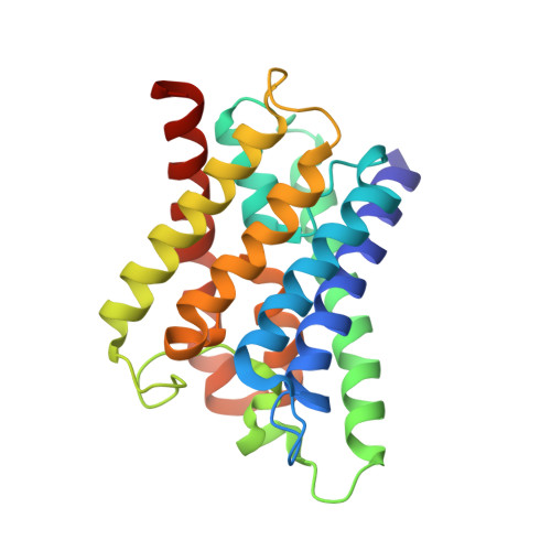
    AqpZ is a homotetramer of four water-conducting channels that facilitate rapid water movements across the plasma membrane of Escherichia coli. Here we report a 3.2 angstroms crystal structure of the tetrameric AqpZ (tAqpZ). All channel-lining residues in the four monomeric channels are found orientated in nearly identical positions with one marked exception at the narrowest channel constriction, where the side chain of a highly conserved Arg-189 adopts two distinct conformational orientations. In one of the four monomers, the guanidino group of Arg-189 points toward the periplasmic vestibule, opening up the constriction to accommodate the binding of a water molecule through a tridentate H-bond. In the other three monomers, the Arg-189 guanidino group bends over to form an H-bond with carbonyl oxygen of the Thr-183, thus occluding the channel. Therefore, the tAqpZ structure reveals two distinct Arg-189 confirmations associated with water permeation through the channel constrictions. Alternation between the two Arg-189 conformations disrupts continuous flow of water, thus regulating the open probability of the water pore. Further, the difference in Arg-189 displacements is correlated with a strong electron density found between the first transmembrane helices of two open channels, suggesting that the observed Arg-189 conformations are stabilized by asymmetrical subunit interactions in tAqpZ.


  • Organizational Affiliation

    Department of Biology, Brookhaven National Laboratory, Upton, New York 11973, USA.


Macromolecules
Find similar proteins by:  (by identity cutoff)  |  3D Structure
Entity ID: 1
MoleculeChains Sequence LengthOrganismDetailsImage
Aquaporin Z
A, B, C, D, E
A, B, C, D, E, F, G, H
231Escherichia coliMutation(s): 0 
Gene Names: aqpZbniP
Membrane Entity: Yes 
UniProt
Find proteins for P60844 (Escherichia coli (strain K12))
Explore P60844 
Go to UniProtKB:  P60844
Entity Groups  
Sequence Clusters30% Identity50% Identity70% Identity90% Identity95% Identity100% Identity
UniProt GroupP60844
Sequence Annotations
Expand
  • Reference Sequence
Small Molecules
Ligands 6 Unique
IDChains Name / Formula / InChI Key2D Diagram3D Interactions
PEE
Query on PEE

Download Ideal Coordinates CCD File 
K [auth A]
O [auth B]
P [auth B]
U [auth E]
W [auth F]
K [auth A],
O [auth B],
P [auth B],
U [auth E],
W [auth F],
X [auth F],
Z [auth G]
1,2-dioleoyl-sn-glycero-3-phosphoethanolamine
C41 H78 N O8 P
MWRBNPKJOOWZPW-NYVOMTAGSA-N
POQ
Query on POQ

Download Ideal Coordinates CCD File 
J [auth A]BIS(((3S,4S,5R,6R)-5-(ETHYL(PHOSPHORYLOXY))-3,4,6-TRIHYDROXY-TETRAHYDRO-2H-PYRAN-2-YL)METHYL) HYDROGEN PHOSPHATE
C16 H33 O20 P3
QTCHWELPTUAUQX-ASCZGESISA-N
AGA
Query on AGA

Download Ideal Coordinates CCD File 
BA [auth G],
S [auth C]
(1S)-2-{[{[(2S)-2,3-DIHYDROXYPROPYL]OXY}(HYDROXY)PHOSPHORYL]OXY}-1-[(PENTANOYLOXY)METHYL]ETHYL OCTANOATE
C19 H36 O10 P
UQSXQYRZHMGKIE-DLBZAZTESA-M
BGL
Query on BGL

Download Ideal Coordinates CCD File 
I [auth A],
L [auth B],
M [auth B],
T [auth E]
2-O-octyl-beta-D-glucopyranose
C14 H28 O6
BVHPDIWLWHHJPD-RKQHYHRCSA-N
3PG
Query on 3PG

Download Ideal Coordinates CCD File 
AA [auth G],
R [auth C]
3-PHOSPHOGLYCERIC ACID
C3 H7 O7 P
OSJPPGNTCRNQQC-UWTATZPHSA-N
PO4
Query on PO4

Download Ideal Coordinates CCD File 
N [auth B],
Q [auth C],
V [auth F],
Y [auth G]
PHOSPHATE ION
O4 P
NBIIXXVUZAFLBC-UHFFFAOYSA-K
Experimental Data & Validation

Experimental Data

  • Method: X-RAY DIFFRACTION
  • Resolution: 3.20 Å
  • R-Value Free: 0.242 
  • R-Value Work: 0.186 
  • R-Value Observed: 0.186 
  • Space Group: P 41 2 2
Unit Cell:
Length ( Å )Angle ( ˚ )
a = 119.152α = 90
b = 119.152β = 90
c = 380.394γ = 90
Software Package:
Software NamePurpose
CNSrefinement
HKL-2000data reduction
SCALEPACKdata scaling
CNSphasing

Structure Validation

View Full Validation Report


Ligand Structure Quality Assessment 


Entry History 

Deposition Data

Revision History  (Full details and data files)

  • Version 1.0: 2005-09-20
    Type: Initial release
  • Version 1.1: 2008-04-30
    Changes: Version format compliance
  • Version 1.2: 2011-07-13
    Changes: Version format compliance
  • Version 1.3: 2014-10-29
    Changes: Non-polymer description
  • Version 1.4: 2020-07-29
    Type: Remediation
    Reason: Carbohydrate remediation
    Changes: Advisory, Data collection, Derived calculations, Structure summary
  • Version 2.0: 2023-08-23
    Changes: Advisory, Atomic model, Data collection, Database references, Derived calculations, Non-polymer description, Refinement description, Structure summary