2A87

Crystal Structure of M. tuberculosis Thioredoxin reductase


Experimental Data Snapshot

  • Method: X-RAY DIFFRACTION
  • Resolution: 3.00 Å
  • R-Value Free: 0.291 
  • R-Value Work: 0.211 
  • R-Value Observed: 0.215 

wwPDB Validation   3D Report Full Report


Ligand Structure Quality Assessment 


This is version 1.3 of the entry. See complete history


Literature

Conformational flexibility of Mycobacterium tuberculosis thioredoxin reductase: crystal structure and normal-mode analysis.

Akif, M.Suhre, K.Verma, C.Mande, S.C.

(2005) Acta Crystallogr D Biol Crystallogr 61: 1603-1611

  • DOI: https://doi.org/10.1107/S0907444905030519
  • Primary Citation of Related Structures:  
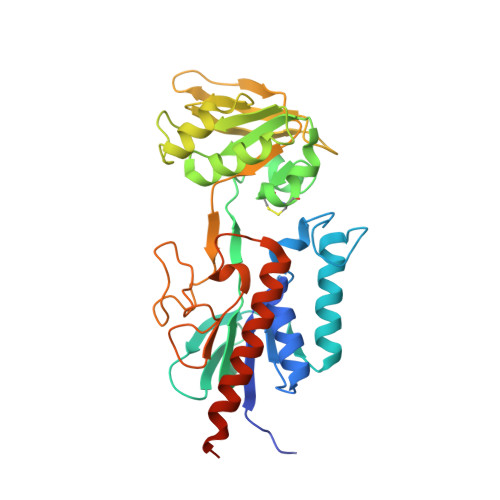
    2A87

  • PubMed Abstract: 

    The thioredoxin system exists ubiquitously and participates in essential antioxidant and redox-regulation processes via a pair of conserved cysteine residues. In Mycobacterium tuberculosis, which lacks a genuine glutathione system, the thioredoxin system provides reducing equivalents inside the cell. The three-dimensional structure of thioredoxin reductase from M. tuberculosis has been determined at 3 A resolution. TLS refinement reveals a large libration axis, showing that NADPH-binding domain has large anisotropic disorder. The relative rotation of the NADPH domain with respect to the FAD domain is necessary for the thioredoxin reduction cycle, as it brings the spatially distant reacting sites close together. Normal-mode analysis carried out based on the elastic network model shows that the motion required to bring about the functional conformational change can be accounted for by motion along one single mode. TLS refinement and normal-mode analysis thus enhance our understanding of the associated conformational changes.


  • Organizational Affiliation

    Centre for DNA Fingerprinting and Diagnostics, Nacharam, Hyderabad, India.


Macromolecules
Find similar proteins by:  (by identity cutoff)  |  3D Structure
Entity ID: 1
MoleculeChains Sequence LengthOrganismDetailsImage
Thioredoxin reductase
A, B
335Mycobacterium tuberculosisMutation(s): 0 
Gene Names: trxB
EC: 1.8.1.9
UniProt
Find proteins for P9WHH1 (Mycobacterium tuberculosis (strain ATCC 25618 / H37Rv))
Explore P9WHH1 
Go to UniProtKB:  P9WHH1
Entity Groups  
Sequence Clusters30% Identity50% Identity70% Identity90% Identity95% Identity100% Identity
UniProt GroupP9WHH1
Sequence Annotations
Expand
  • Reference Sequence
Experimental Data & Validation

Experimental Data

  • Method: X-RAY DIFFRACTION
  • Resolution: 3.00 Å
  • R-Value Free: 0.291 
  • R-Value Work: 0.211 
  • R-Value Observed: 0.215 
  • Space Group: P 41 21 2
Unit Cell:
Length ( Å )Angle ( ˚ )
a = 107.4α = 90
b = 107.4β = 90
c = 118.2γ = 90
Software Package:
Software NamePurpose
REFMACrefinement
DENZOdata reduction
SCALEPACKdata scaling
AMoREphasing

Structure Validation

View Full Validation Report


Ligand Structure Quality Assessment 


Entry History 

Deposition Data

Revision History  (Full details and data files)

  • Version 1.0: 2005-11-29
    Type: Initial release
  • Version 1.1: 2008-04-30
    Changes: Version format compliance
  • Version 1.2: 2011-07-13
    Changes: Advisory, Version format compliance
  • Version 1.3: 2023-08-23
    Changes: Data collection, Database references, Derived calculations, Refinement description