1ZX4

Structure of ParB bound to DNA


Experimental Data Snapshot

  • Method: X-RAY DIFFRACTION
  • Resolution: 2.98 Å
  • R-Value Free: 0.296 
  • R-Value Work: 0.248 
  • R-Value Observed: 0.248 

wwPDB Validation   3D Report Full Report


This is version 1.3 of the entry. See complete history


Literature

Structures of ParB bound to DNA reveal mechanism of partition complex formation.

Schumacher, M.A.Funnell, B.E.

(2005) Nature 438: 516-519

  • DOI: https://doi.org/10.1038/nature04149
  • Primary Citation of Related Structures:  
    1ZX4

  • PubMed Abstract: 

    The faithful inheritance of genetic information, which is essential for all organisms, requires accurate DNA partition (segregation) at cell division. In prokaryotes, partition is mediated by par systems, for which the P1 plasmid system of Escherichia coli is a prototype comprising a partition site and two proteins, ParA and ParB. To form the partition complex necessary for segregation, P1 ParB must recognize a complicated arrangement of A-box and B-box DNA motifs located on opposite ends of a sharply bent parS partition site of approximately 74 bp (refs 3-7). Here we describe structures of ParB bound to partition sites. ParB forms an asymmetric dimer with extended amino-terminal HTH (helix-turn-helix) domains that contact A-boxes. The two HTH domains emanate from a dimerized DNA-binding module composed of a six-stranded beta-sheet coiled-coil that binds B-boxes. Strikingly, these individual DNA-binding modules rotate freely about a flexible linker, enabling them to contact several arrangements of A- and B-boxes. Most notably, each DNA-binding element binds to and thus bridges adjacent DNA duplexes. These unique structural features of ParB explain how this protein can bind complex arrays of A- and B-box elements on adjacent DNA arms of the looped partition site.


  • Organizational Affiliation

    Department of Biochemistry and Molecular Biology, Oregon Health & Science University, Portland, Oregon 97239-3098, USA. schumacm@ohsu.edu


Macromolecules

Find similar proteins by:  (by identity cutoff)  |  3D Structure
Entity ID: 3
MoleculeChains Sequence LengthOrganismDetailsImage
Plasmid Partition par B proteinC [auth A],
D [auth B]
192Punavirus P1Mutation(s): 6 
Gene Names: parb
UniProt
Find proteins for Q38420 (Punavirus P1)
Explore Q38420 
Go to UniProtKB:  Q38420
Entity Groups  
Sequence Clusters30% Identity50% Identity70% Identity90% Identity95% Identity100% Identity
UniProt GroupQ38420
Sequence Annotations
Expand
  • Reference Sequence

Find similar nucleic acids by:  Sequence   |   3D Structure  

Entity ID: 1
MoleculeChains LengthOrganismImage
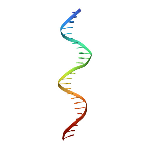
parS-small DNA centromere siteA [auth T]25N/A
Sequence Annotations
Expand
  • Reference Sequence

Find similar nucleic acids by:  Sequence   |   3D Structure  

Entity ID: 2
MoleculeChains LengthOrganismImage
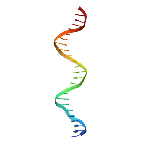
parS-small DNA centromere siteB [auth S]25N/A
Sequence Annotations
Expand
  • Reference Sequence
Small Molecules
Modified Residues  1 Unique
IDChains TypeFormula2D DiagramParent
MSE
Query on MSE
C [auth A],
D [auth B]
L-PEPTIDE LINKINGC5 H11 N O2 SeMET
Experimental Data & Validation

Experimental Data

  • Method: X-RAY DIFFRACTION
  • Resolution: 2.98 Å
  • R-Value Free: 0.296 
  • R-Value Work: 0.248 
  • R-Value Observed: 0.248 
  • Space Group: P 62 2 2
Unit Cell:
Length ( Å )Angle ( ˚ )
a = 154.6α = 90
b = 154.6β = 90
c = 132.3γ = 120
Software Package:
Software NamePurpose
MOSFLMdata reduction
SCALAdata scaling
SOLVEphasing
CNSrefinement
CCP4data scaling

Structure Validation

View Full Validation Report


Entry History 

Deposition Data

Revision History  (Full details and data files)

  • Version 1.0: 2005-11-29
    Type: Initial release
  • Version 1.1: 2008-04-30
    Changes: Version format compliance
  • Version 1.2: 2011-07-13
    Changes: Version format compliance
  • Version 1.3: 2017-10-04
    Changes: Structure summary